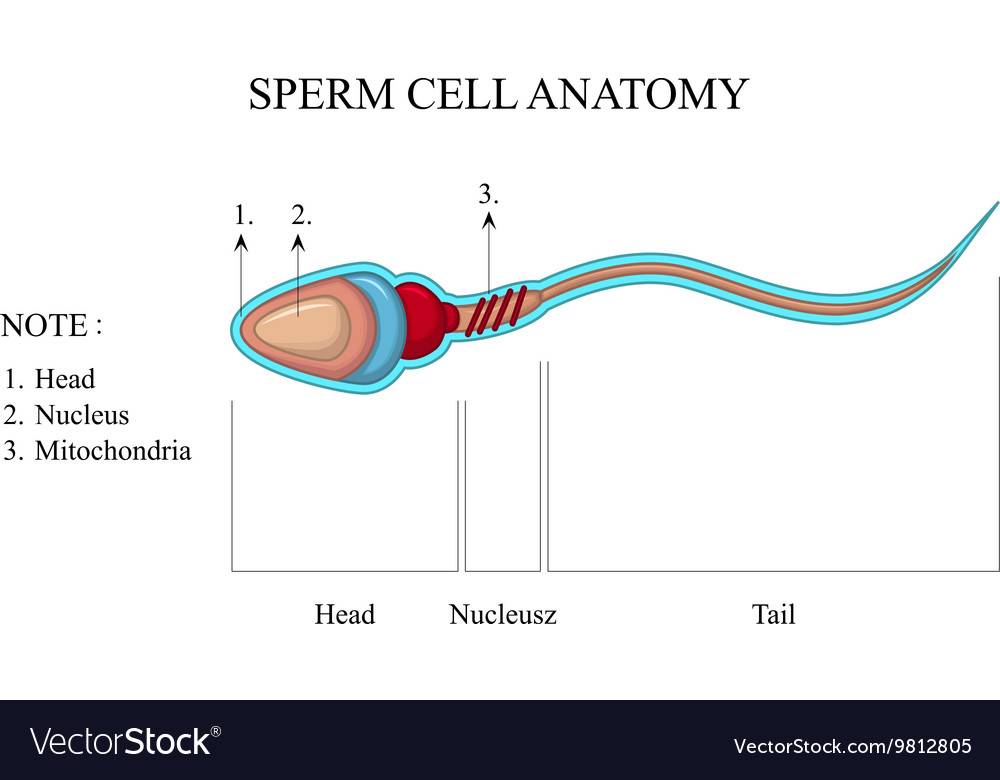
Sperm cell anatomy, education fertility diagram VectorMine

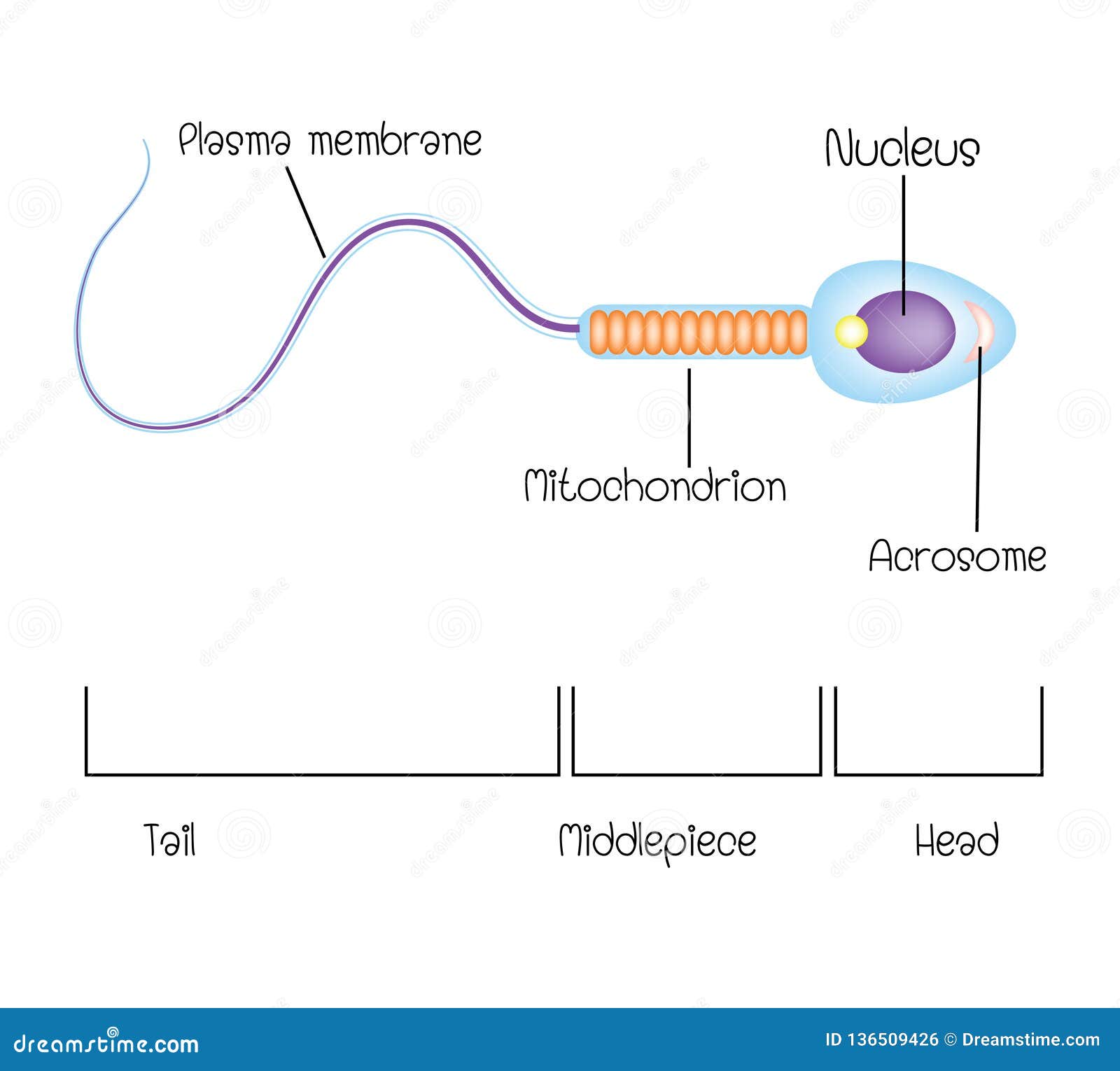
Draw a labelled diagram of the microscopic structure of a human sperm
Spermatogenesis. Spermatogenesis is the process of formation of mature sperm cells through a series of mitotic and meiotic divisions along with metamorphic changes in the immature sperm cell.. It is the male version of gametogenesis which results in the formation of mature male gametes. In mammals, this takes place in the seminiferous tubules of the male reproductive system.
How is the body of a sperm suited for fertilization of an egg? Socratic
Structure of sperm cell, how to draw the sperm cell easily and simply, sperm cell diagram with in 5 minutes, colour full sperm cell diagram, how to draw th.

Cell structure — the science hive
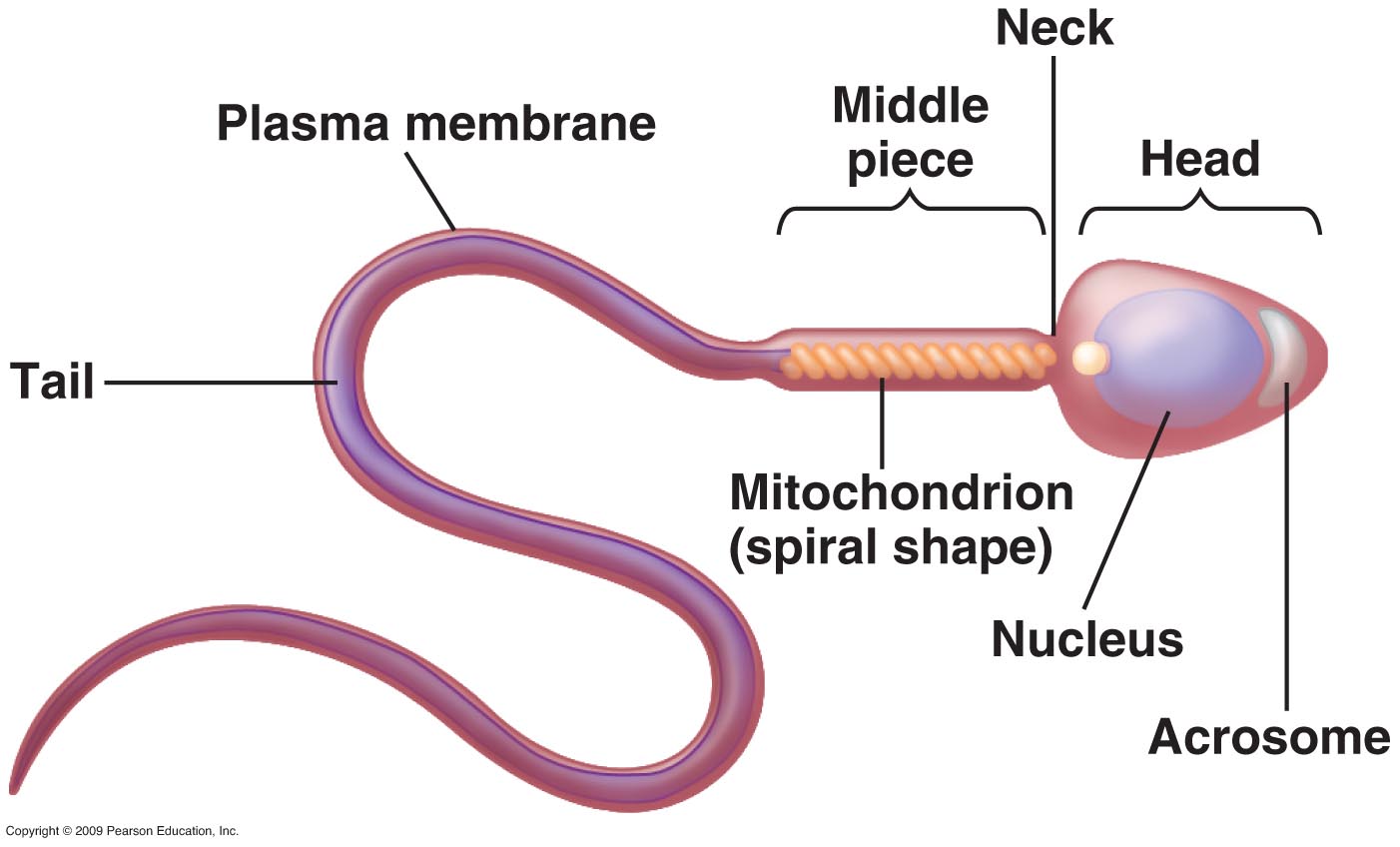
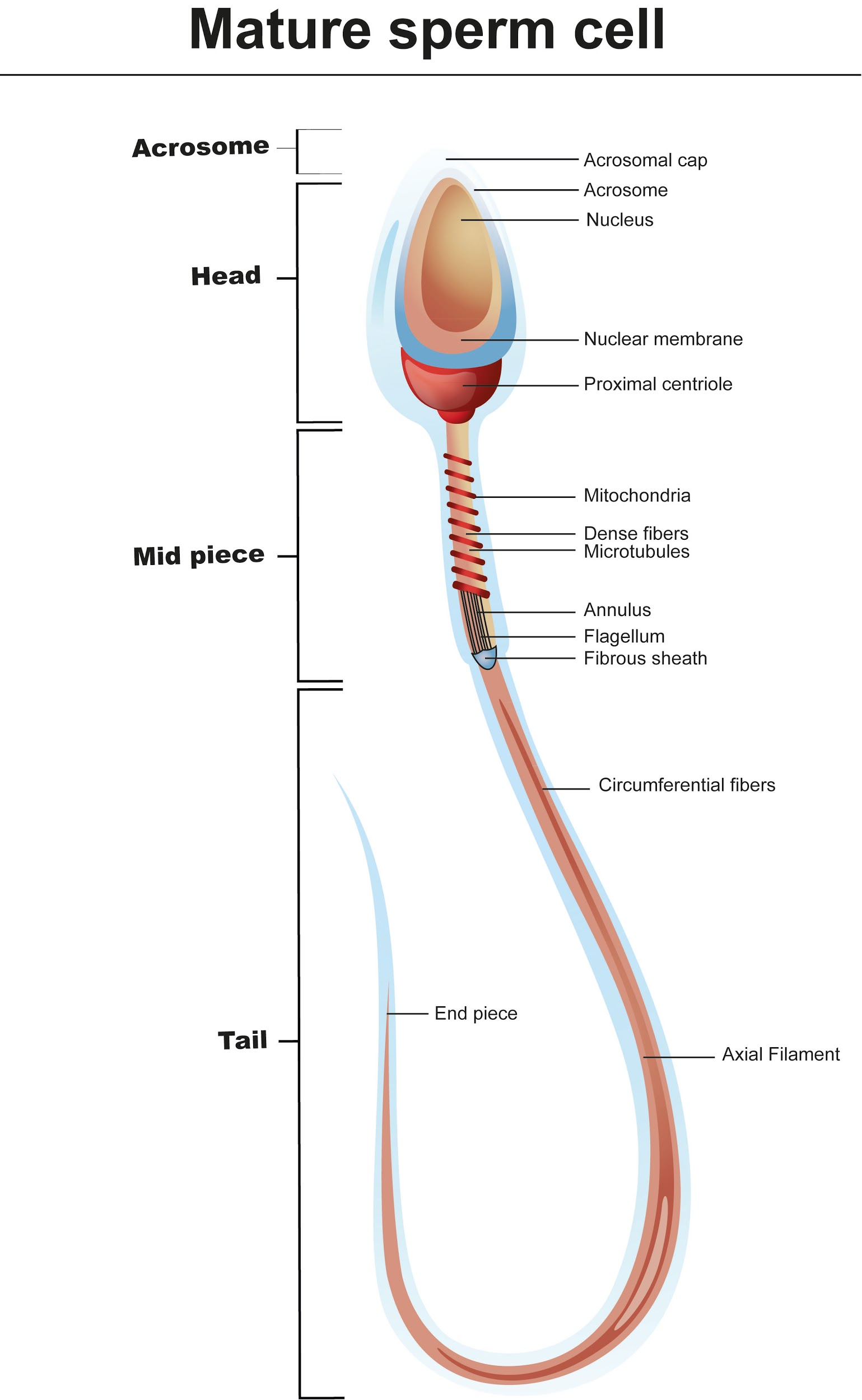
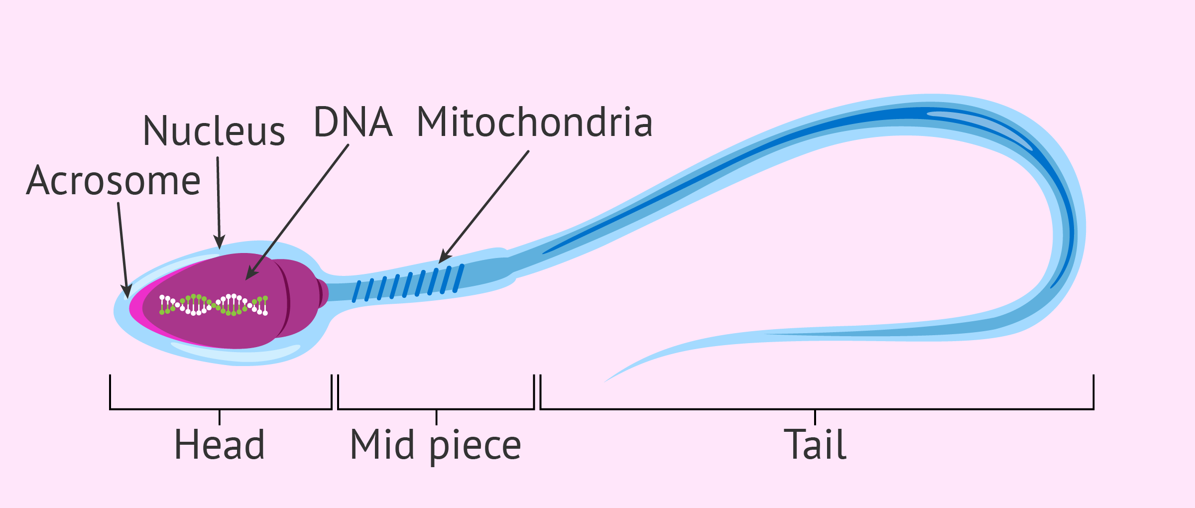
A mature sperm cell has several structures that help it reach and penetrate an egg. These are labeled in the drawing of a sperm shown in Figure \(\PageIndex{2}\). The head is the part of the sperm that contains the nucleus — and not much else. The nucleus, in turn, contains tightly coiled DNA that is the male parent's contribution to the.
Draw the diagram of the human sperm and label its parts. Describe it in
lysin See all related content → sperm, male reproductive cell, produced by most animals. With the exception of nematode worms, decapods (e.g., crayfish), diplopods (e.g., millipedes), and mites, sperm are flagellated; that is, they have a whiplike tail. In higher vertebrates, especially mammals, sperm are produced in the testes.
We Love Cellz ★
Definition: What are Sperm Cells? Sperm cells are gametes (sex cells) that are produced in the testicular organ (gonad) of male human beings and animals. Like the female gamete (oocyte), sperm cells carry a total of 23 chromosomes that are a result of a process known as meiosis.
Biology Cell Specialisation HubPages
It carries and stores the sperm cells that your testicles create. The epididymis also brings the sperm to maturity — the sperm that emerge from the testicles are immature and incapable of fertilization. During sexual arousal, muscle contractions force the sperm into the vas deferens. What are the internal parts of the male reproductive system?
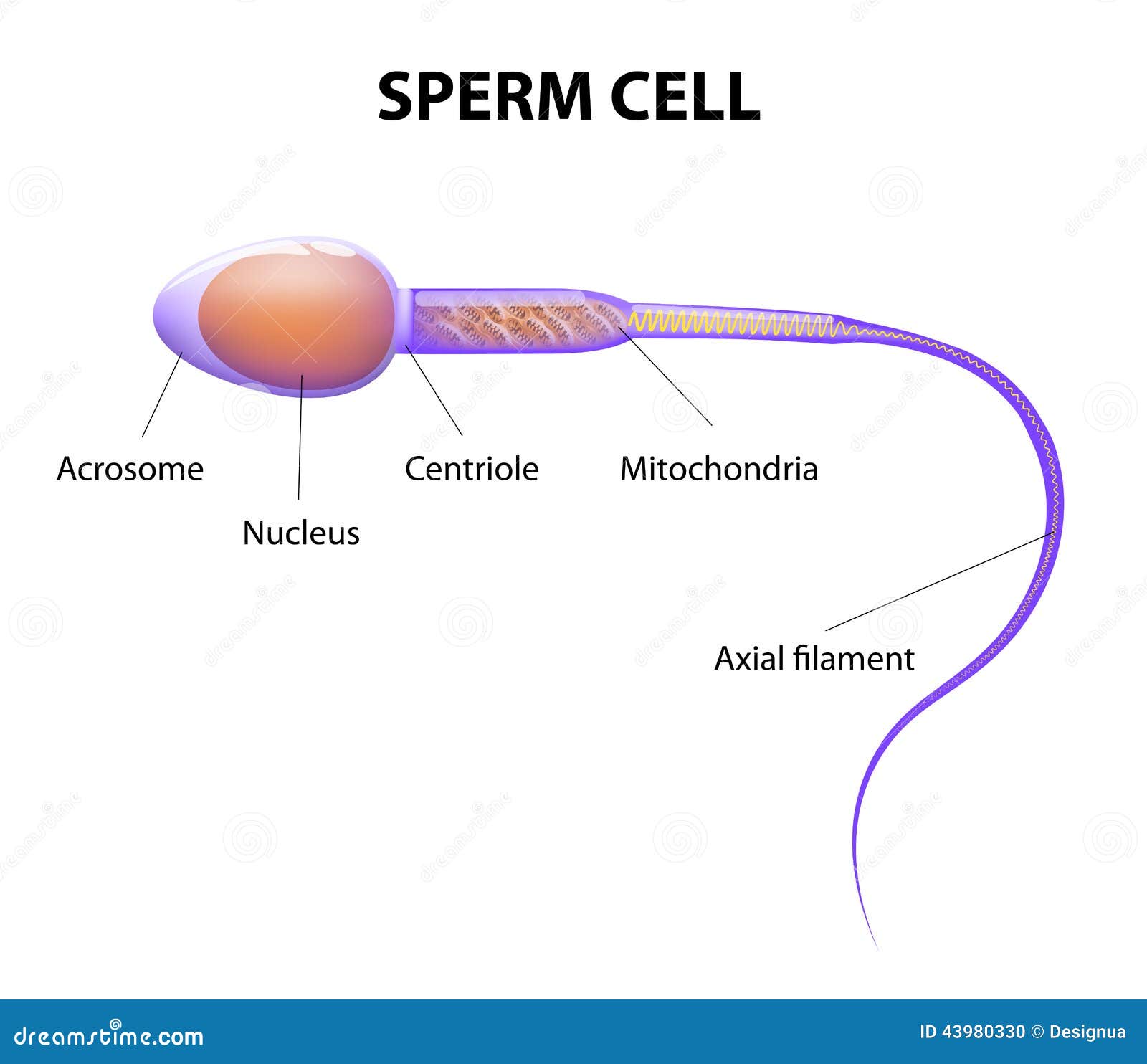
Structure Of A Sperm Cell Stock Vector Image 43980330
A spermatozoon, in plural spermatozoa, or sperm cell is the male reproductive cell that is produced in the man´s testicles in a process called spermatogenesis. The sperm cell´s function is to enable sexual reproduction through its union with the female egg during fertilization.
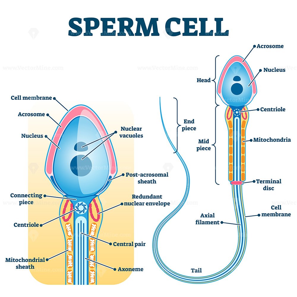
Sperm cell anatomy, education fertility diagram VectorMine
This labelled diagram shows the structure of a sperm cell in detail, which has the following parts: Head With its spheric shape, it consists of a large nucleus, which at the same time contains an acrosome. The nucleus contains the genetic information and 23 chromosomes.
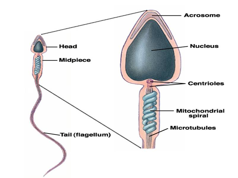
The middle piece of the sperm contains(a)Proteins(b)Centriole(c)Nucleus
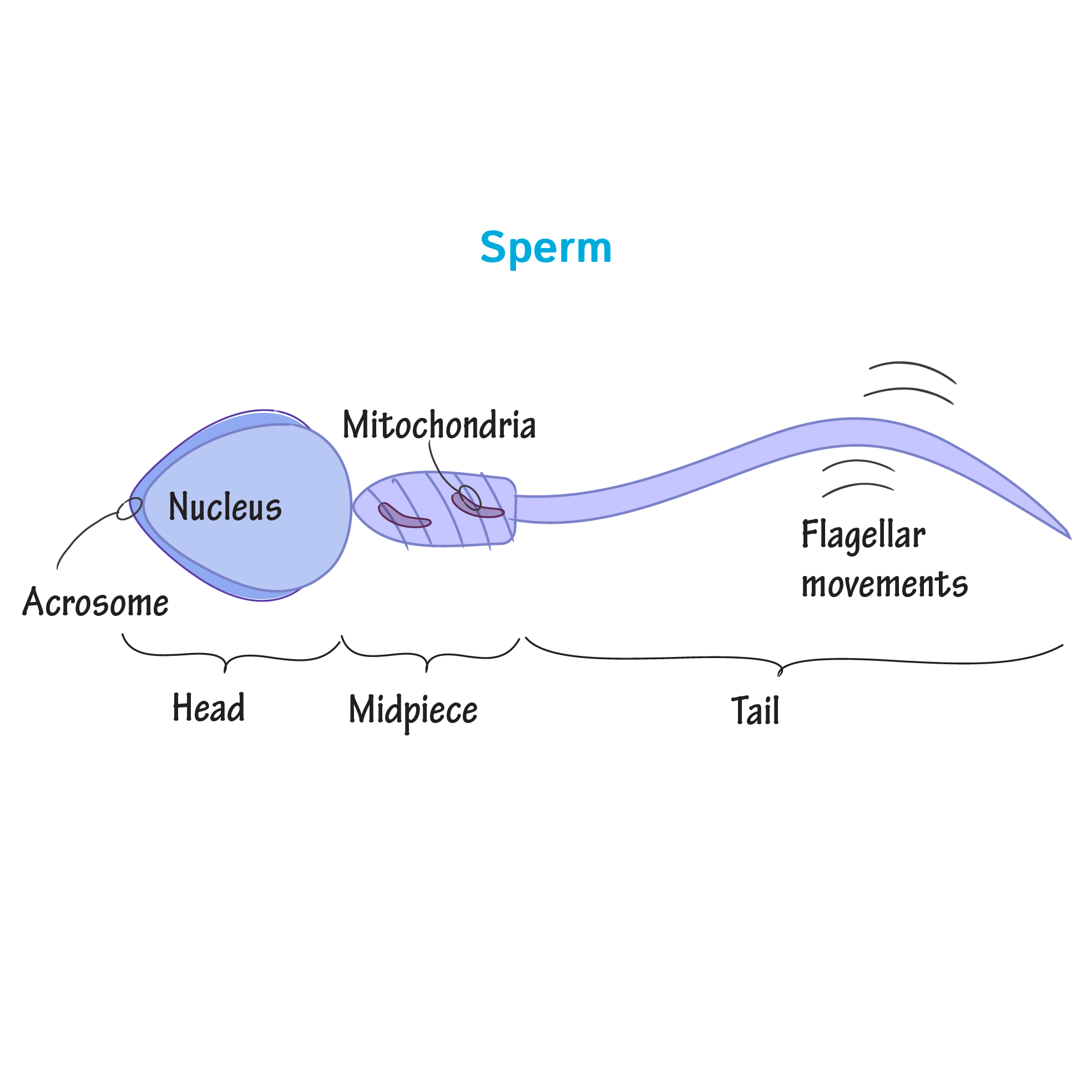
The sperm cell diagram below shows multiflagellate fern cells. Sperm cells from the fern plant. Most motile spermatozoa have flagella to help them swim through fluids - the seminal fluid produced by males and the mucus membranes of the female reproductive tract. Flagellum movement requires a consistent energy source.
Sperm Anatomy
Diagram of a human sperm cell Sperm ( pl.: sperm or sperms) is the male reproductive cell, or gamete, in anisogamous forms of sexual reproduction (forms in which there is a larger, female reproductive cell and a smaller, male one).

Draw a diagram of the microscopic structure of human sperm. Label the
spermatozoonɜːr [1] also spelled spermatozoönspermatozoa; from Ancient Greek σπέρμα spérma 'seed', and ζῷον zôion 'animal') is a , or moving form of the haploid that is the male gamete. A spermatozoon joins ovum to form a zygote. (A zygote is a single cell, with a complete set of chromosomes, that normally develops into an embryo .)

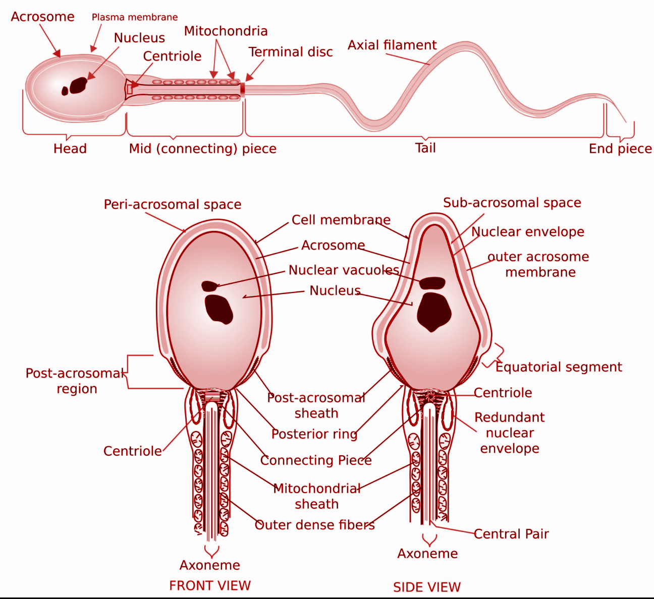
Structural sperm features. Spermatozoa are composed of two main
Sperm is the male reproductive cell or gamete. The term "gamete" implies that the cell is half of a whole. When a sperm combines with a female gamete, or egg, it results in a human embryo.
Structure of a sperm cell stock vector. Illustration of evolution
22.3: Structure of Formed Sperm. Sperm are smaller than most cells in the body; in fact, the volume of a sperm cell is 85,000 times less than that of the female gamete. Approximately 100 to 300 million sperm are produced each day, whereas women typically ovulate only one oocyte per month. As is true for most cells in the body, the structure of.
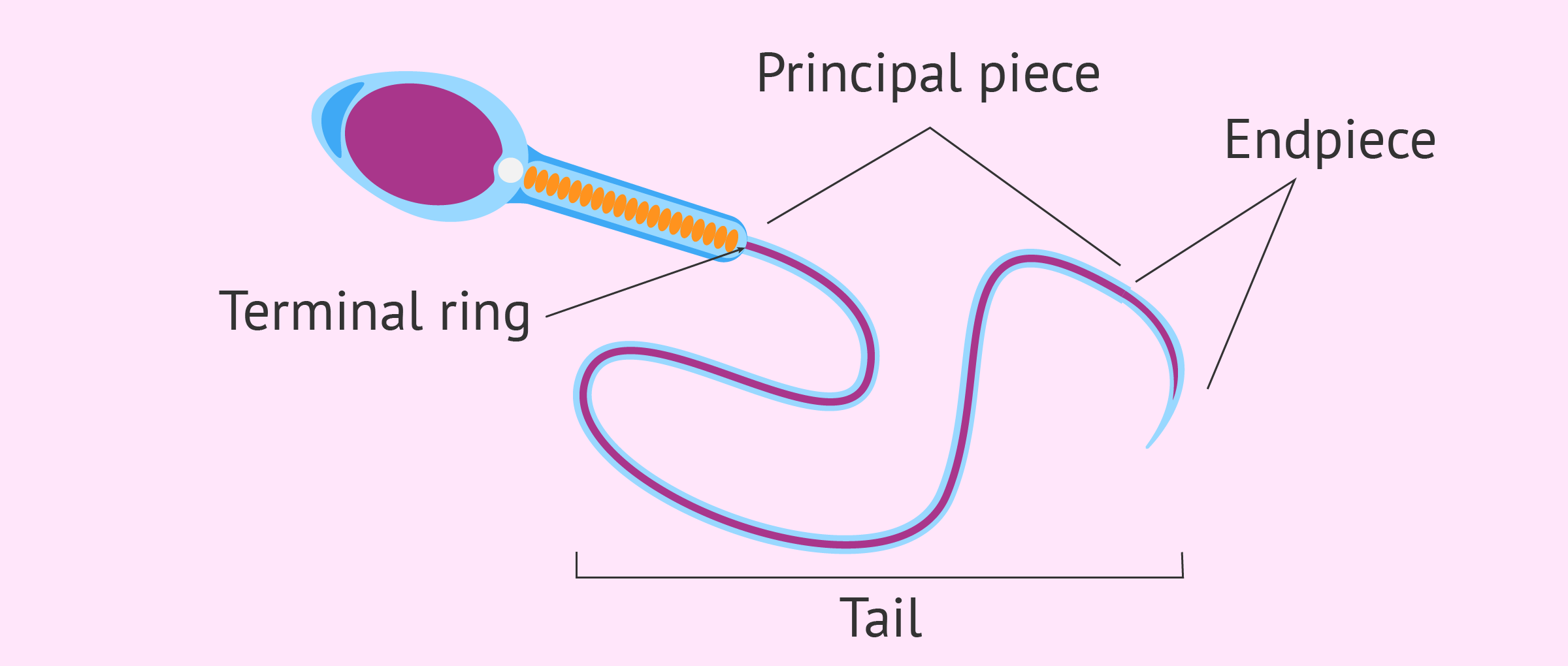
Diagram of sperm cell tail
'How to draw Sperm Cell || Study of Human Spermatozoon diagram and label the parts' is demonstrated in this video tutorial step by step.Sperm is the male rep.
Human sperm cell anatomy Royalty Free Vector Image
Diagram of a sperm cell showing many detailed components Summary [ edit] File history Click on a date/time to view the file as it appeared at that time. You cannot overwrite this file. File usage on Commons The following 26 pages use this file: User:LadyofHats/gallery1 User:Rocket000/SVGs/Biology File:Complete diagram of a human spermatozoa-ar.svg
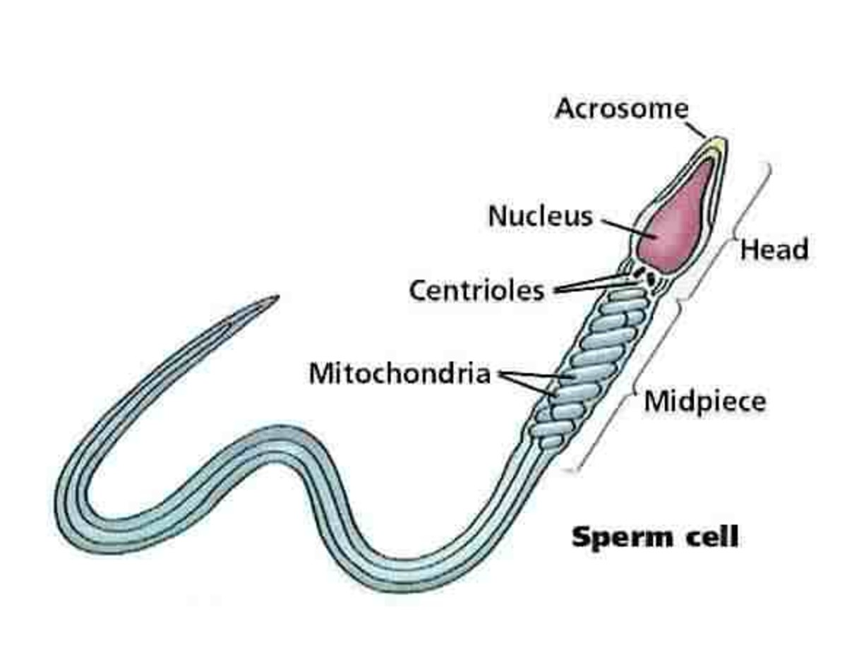
Structure of a mature human sperm cell
Sperm - Molecular Biology of the Cell - NCBI Bookshelf , however, the progeny of a single spermatogonium develop as a large In most species, there are just two types of gamete, and they are radically different. The egg is among the largest cells in an organism, while the sperm (spermatozoon, plural spermatozoa) is often the smallest.
