Diagrams of Microscope 101 Diagrams
MICROSCOPE DIAGRAM Unmasa Dalha
The web page titled "Parts of a Microscope with Labeled Diagram and Functions" has the following key takeaways: 🔍 The microscope is an essential tool for scientists, researchers, and medical professionals. 🧬 The main function of a microscope is to provide a magnified view of small objects or organisms, such as bacteria, cells, or tissues.
Microscope Diagram to Print 101 Diagrams
Drag and drop the text labels onto the microscope diagram. If you want to redo an answer, click on the box and the answer will go back to the top so you can move it to another box. If you want to check your answers, use the Reset incorrect button. This will reset incorrect answers only.
1.5 Microscopy Biology LibreTexts
With Labeled Diagram and Functions How does a Compound Microscope Work? Before exploring microscope parts and functions, you should probably understand that the compound light microscope is more complicated than just a microscope with more than one lens.
Ag Biology Unit 2
A Study of the Microscope and its Functions With a Labeled Diagram To better understand the structure and function of a microscope, we need to take a look at the labeled microscope diagrams of the compound and electron microscope. These diagrams clearly explain the functioning of the microscopes along with their respective parts.
Light Microscope Main Parts Of Light Microscope Biology —
Microscope - Types, Diagrams and Functions By Editorial Board October 13, 2022 Microscope - Let's split the name into two parts to understand what it actually means. " Micro " means very small (typically not visible to the naked eye) and " scope " means to assess or investigate carefully.

Light Microscope Definition, Principle, Types, Parts, Labeled Diagram
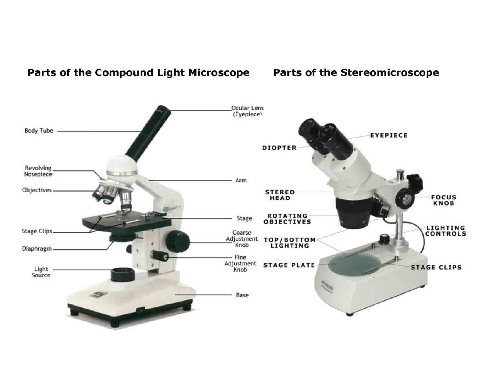
Parts of the Microscope (Labeled Diagrams) By Editorial Board December 14, 2022 The microscope is one of the must-have laboratory tools because of its ability to observe minute objects, usually living organisms that cannot be seen by the naked eyes. It is categorized into two: simple and compound microscopes.
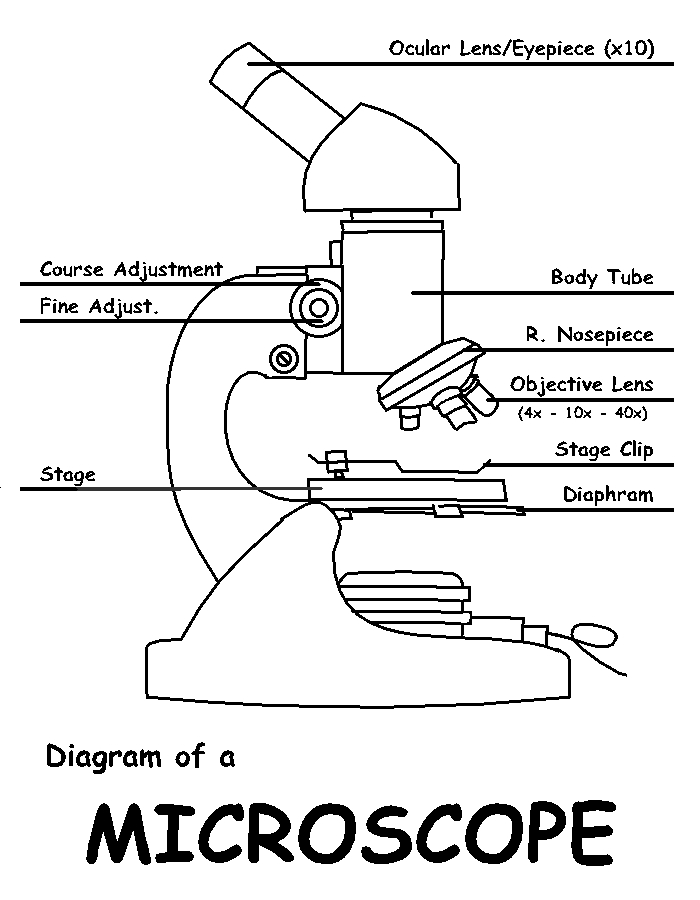
Diagram of a Microscope by ScienceDoodles on DeviantArt
Q. Diagrammatically, identify the various parts of a microscope. Q. Describe the functions of each part of the microscope you have drawn above. Q. Differentiate between a condenser and an Abbe condenser. Q. What is the magnification power of the objective lenses? Q. How does the eyepiece compare to the objective lens? Q.

Microscope Drawing And Label at GetDrawings Free download
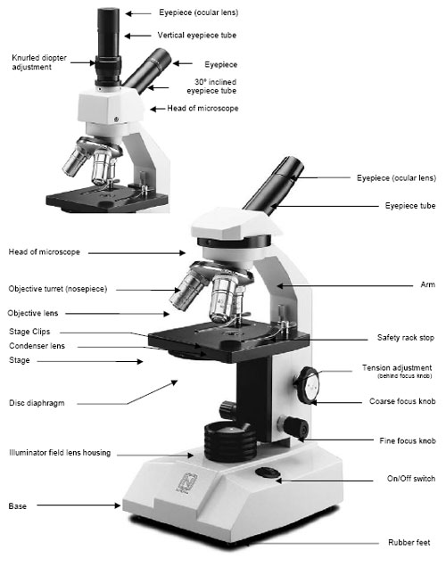
The field diaphragm control is located around the lens located in the base. Hinge Screw -This screw fixes the arm to the base and allow for the tilting of the arm. Stage Clips - They hold the slide firmly onto the stage. On/OFF Switch - This switch on the base of the microscope turns the illuminator off and on.

36 Label Parts Of The Microscope Labels 2021
There are 1000 millimeters (mm) in one meter. 1 mm = 10 -3 meter. There are 1000 micrometers (microns, or µm) in one millimeter. 1 µm = 10 -6 meter. There are 1000 nanometers in one micrometer. 1 nm = 10 -9 meter. Figure 1: Resolving Power of Microscopes. The microscope is one of the microbiologist's greatest tools.
Microscope Parts Sketch at Explore collection of
This activity has been designed for use in homes and schools. Each microscope layout (both blank and the version with answers) are available as PDF downloads. You can view a more in-depth review of each part of the microscope here. Download the Label the Parts of the Microscope PDF printable version here.

Microscope labeled diagram
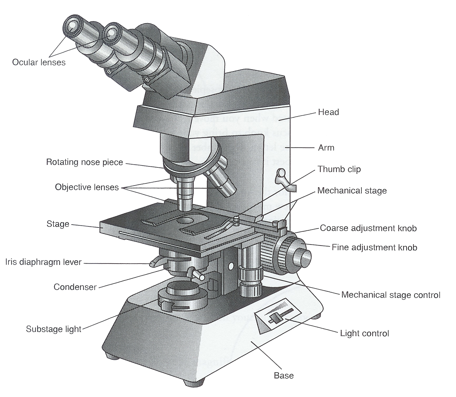
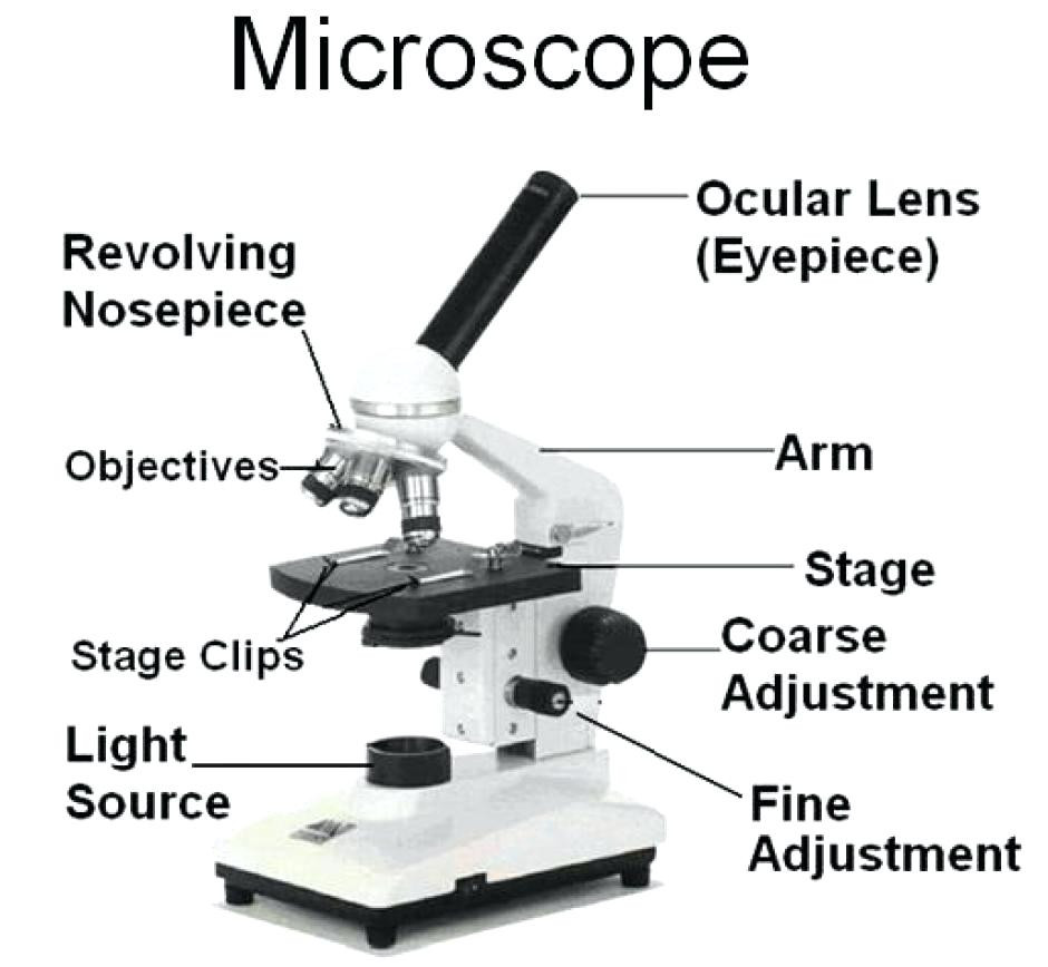
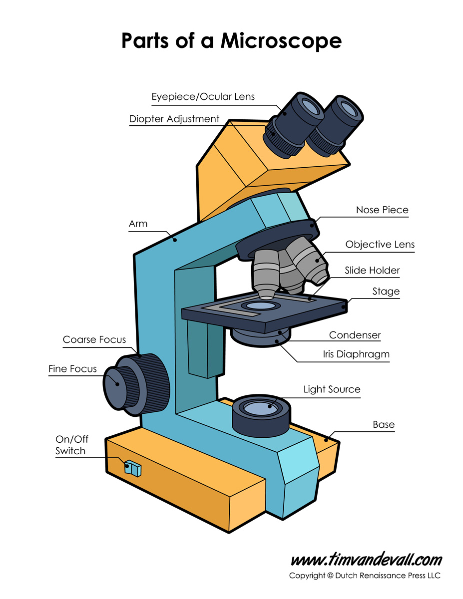
1. Eyepiece 2. Body tube/Head 3. Turret/Nose piece 4. Objective lenses 5. Knobs (fine and coarse) 6. Stage and stage clips 7. Aperture 9. Condenser 10. Condenser focus knob 11. Iris diaphragm 12. Diopter adjustment 13. Arm 14. Specimen/slide 15. Stage control/stage height adjustment 16. On and off switch 17. Base
Monday September 25 Parts of a Compound Light Microscope
Diaphragm (Iris) Condenser Aperture Stage Objective lens Body Tube Ocular Lens (eye-piece) Coarse and Fine Adjustment Knob Arm Base Microscope Worksheet The Light Microscope Light microscopes are used to examine cells at relatively low magnifications. Magnifications of about 2000X are the upper limit for light microscopes.

Microscope diagram Tom Butler Technical Drawing and Illustration
Labeled parts of a microscope. General Rules. Always START and END with the low power lens when putting on OR taking away a slide. Never turn the nose piece by the objective lens. Do not get any portion of the microscope wet - especially the stage and objective lenses.

Microscope Diagram Labeled, Unlabeled and Blank Parts of a Microscope
First and foremost, we have a labeled microscope diagram, available in both black and white and color. Useful as a study guide for learning the anatomy of a microscope. There are six printables available. You can download them individually by clicking the images below, or download them together in a single pdf bundle here. [clearBoth]
How to Use a Microscope
What Is a Microscope? are instruments designed to produce magnified visual or photographic images of small objects. A microscope must accomplish three tasks: produce a magnified image, separate the details in the image, and render these details visible to the human eye or camera.

Microscope Diagram to Print 101 Diagrams
Figure: Labeled Diagram of a Light Microscope. Types of light microscopes (optical microscope) With the evolved field of Microbiology, the microscopes. used to view specimens are both simple and compound light microscopes, all using lenses. The difference is simple light microscopes use a single lens for magnification while compound lenses use.
