Muscle Anatomy Wall Charts Set Of Muscle Anatomy Anatomy Human My XXX

Anatomy Art Print Set 6 Medical Wall Art Anatomical Poster Etsy
In this article, we shall look at the anatomy of the anterolateral abdominal wall - its musculature, surface anatomy and clinical correlations. Superficial Fascia The superficial fascia is connective tissue . The composition of this layer depends on its location: Above the umbilicus - a single sheet of connective tissue.

Human Anatomy Print Set of 6, Medical Office Wall Art Decor, Vintage
The abdominal wall surrounds the anterolateral aspect of the abdominal cavity, where many important organs are located. [1] [2] [3] Chief layers of the abdominal wall include: Skin, Superficial fascia (the subcutaneous tissue which forms the thin, single layer above the umbilicus.
The Anatomy of an Interior Wall The Graceful Dwelling
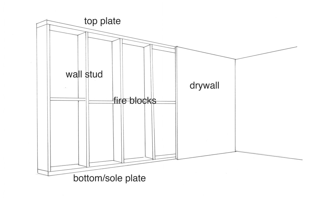
Our anatomy of a wall illustration below is split into 3 sections. Those sections, from top to bottom, are: Wall exterior; Wall interior (i.e. the stud or framed wall); and Typical wall layers (exterior wall). Diagram: Anatomy of a Wall Wall Surface
Vintage Anatomy Wall Chart of the Upper Body, circa 1960 For Sale at
RENT "ANATOMY OF A FALL" ON PRIME VIDEO $5.99. A courtroom drama exploring the collapse of a marriage, the film stars Sandra Huller ("The Zone of Interest") as a novelist who is put on.

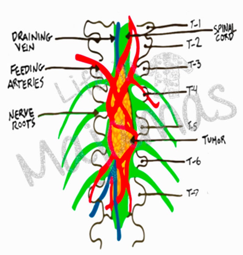
Figure 4 from Introduction to chest wall reconstruction anatomy and
Anatomy of the anterolateral abdominal wall Figure 1: Muscles of the anterolateral abdominal wall A. Superficial B. Deeper dissection. Figure 2: Layers of the anterolateral abdominal wall. Figure 3: Rectus sheath. A. Sagittal view. B. Posterior view of anterior abdominal wall.
Traumatic Hemothorax Core EM
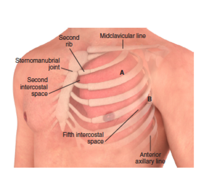
Thoracic wall The first step in understanding thorax anatomy is to find out its boundaries. The thoracic, or chest wall, consists of a skeletal framework, fascia, muscles, and neurovasculature - all connected together to form a strong and protective yet flexible cage.

Buy Spectrum Pre School Kids Learning Educational Human Anatomy Name
Fig. 2.27. The transversus muscle is the deepest of the anterolateral abdominal wall muscles; it arises from the iliopsoas fascia and inner lip of the iliac crest in its anterior two-thirds. The muscle extends to the inner surfaces of the lowest six costal cartilages, and its aponeurosis extends to the linea alba.
human visceral anatomy
Overview of the surface anatomy landmarks found in the abdomen and lower limbs. Abdomen 1/9 Synonyms: Abdominal region, Regio abdominis , show more. The abdominal wall surrounds the abdominal cavity, providing it with flexible coverage and protecting the internal organs from damage.
Grey's Anatomy Wall Tumor PNG Etsy
Therefore, my official prediction is that "Anatomy of a Fall" should arrive on Hulu for streaming in early April 2024 . There's a chance it comes a bit earlier to try and ride the wave from the.
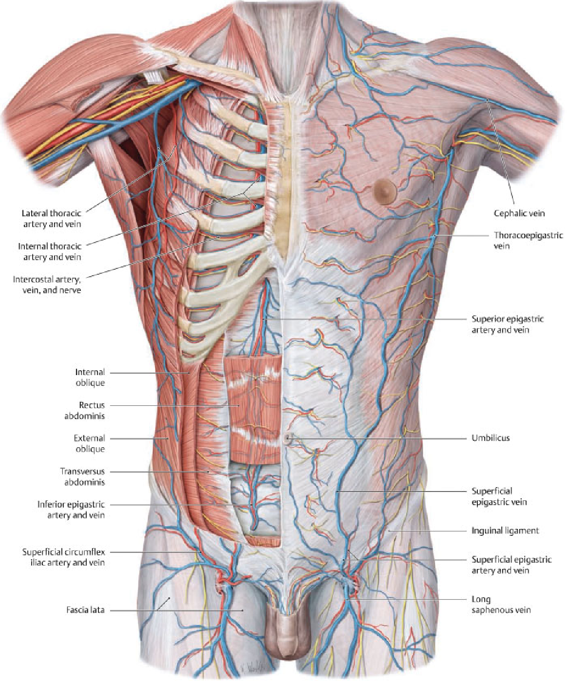
diagram of chest anatomy
Streaming options for all countries explored. After snagging the Palme d'Or at the Cannes Film Festival, the film Anatomy of a Fall has made a big impression on movie screens. Viewers loved the.

Vintage Human Anatomy Prints Set of 6 (8 inches x 10 inches
The abdominal wall is a complex organ with many functions that contribute to a patient's quality of life. The anatomical core of the anterolateral abdominal wall is mainly comprised of 4 paired symmetrical muscles. Classically the anterolateral abdominal wall has been described as separate layers from superficial to deep as follows:

chest wall muscle anatomy
Anatomy of a Wall How Walls Are Built by Don Vandervort, HomeTips © 1997 to 2023 October 25, 2020 Basic interior wall-framing components, including drywall, plaster, and panel constructions In the illustration below, you can see an interior wall's basic wall-framing components.

Human Brain Print Human Anatomy Wall Art Medical Wall Decor Male Figure
An abdominal wall formed of skin, fascia, and muscle encases the abdominal cavity and viscera. The abdominal wall does not only contain and protect the intra-abdominal organs but can distend, generate intrabdominal pressure, and move the vertebral column.

Abdominal Wall Anatomy Of The Abdomen Learn Surgery
The anterior abdominal wall extends from the xiphoid process and. costal margins. cranially to the pubic and iliac bones inferiorly and to the. mid-axillary lines. on either side. The abdomen is divided into regions or quadrants to more precisely describe abdominal symptoms and signs and help identify underlying organs.
368284 HUMAN ANATOMY Interactive Body Wall Chart Art Decor Print Poster
Anatomy of the Inguinocrural ea Ar The surgical space called the Fruchaud triangle is an opening of the lower abdomi-nal wall, bounded by the conjoint tendon, the iliopubiana branch, the rectus abdomi-nis, and the iliopsoas. This triangle is divided by the inguinal ligament into two topographical regions: inguinoabdominal and inguinocrural.

Buy CHDITB Unframed Flowers Skeletal Wall Art Print, Human Organs Art
There are nine layers to the abdominal wall: skin, subcutaneous tissue, superficial fascia, external oblique muscle, internal oblique muscle, transversus abdominis muscle, transversalis fascia, preperitoneal adipose and areolar tissue, and peritoneum. Nerves, blood vessels, and lymphatics are present throughout.
