Imaging of Elbow Fractures and Dislocations in Adults in 2023 Radiology student, Human anatomy

Radiographic Anatomy of the Elbow
The normal carrying angle (5 to 20 degrees, average 15 degrees) can be measured on the AP view. 1,5. FIGURE 6-1 A, Patient positioned for the anteroposterior (AP) view of the elbow. The arm is level with the cassette, with the hand positioned palm up. The central beam (pointer) is perpendicular to the elbow.

Radiographic Anatomy Elbow Oblique Medical knowledge, Radiology, Medical anatomy
Indications This view is clinically indicated for trauma, chronic discomfort or infection of the elbow joint. It aids in visualizing fractures and/or dislocations of the elbow joint, in addition to osteomyelitis and arthritic changes.

Imaging of Elbow Fractures and Dislocations in Adults in 2023 Radiology student, Human anatomy
7.2K Description Labeled Elbow XRay Anatomy - Lateral View #Anatomy #Radiology #Elbow #XRay #Lateral #Labeled Contributed by Dr. Gerald Diaz @ GeraldMD Board Certified Internal Medicine Hospitalist, GrepMed Editor in Chief 🇵🇭 🇺🇸 - Sign up for an account to like, bookmark and upload images to contribute to our community platform.

Lateral Xray of elbow Radiology student, Radiology, Radiologic technology
Edit article Citation, DOI, disclosures and article data The lateral elbow view is part of the two view elbow series, examining the distal humerus, proximal radius and ulna. It is deceptively one of the more technically demanding projections in radiography 1-3.

Labeled elbow Radiographs Radiology, Anatomy, Radiography
The lateral elbow is a troublesome radiographic position in terms of achieving a true lateral view. If you haven't achieved a true lateral view, understanding how to correct the position can also prove difficult.

Labeled elbow Radiographs Jeremy Jones, Radiology Tech, Xray Technician, Radiographer, Rad Tech
Adults: Treat as radial head fracture Peds: Be certain that neither an undisplaced supracondylar fracture nor a displaced internal epicondyle fracture is overlooked! Is the radiocapitellar line normal? A line drawn along the longitudinal axis of the radial head and neck should pass through the capitellum
Elbow Dislocation Core EM
An X-ray of the elbow is a frequently conducted examination and is mainly used for diagnosing a fracture. Some of the key topics are radial head fracture, supracondylar humeral fracture, anterior/posterior fat pad and elbow luxation. Prior to this module, it is wise to read the Fracture General Principles module.

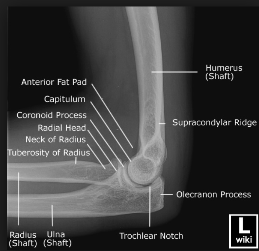
Radiographic Anatomy Elbow Lateral Medical anatomy, Radiology student, Radiology
Oblique Elbow. Humerus (shaft) Lateral supraepicondylar ridge. Lateral epicondyle. Capitellum of humerus. Radial fossa. Medial supraepicondylar ridge. Medial epicondyle. Trochlea of humerus.
EPOS™
This is often the only X-ray sign of a bone injury. A post-traumatic effusion without a visible bone fracture usually indicates a radial head fracture in an adult, and a supracondylar fracture of the distal humerus in a child. If there is a joint effusion but no history of trauma, an inflammatory cause should be considered.
elbow x ray anatomy
Imaging Imaging of the Elbow By admin On Jan 11, 2023 The elbow joint is a complex structure with three separate intracapsular articulations. These are highly congruent joints with multiplanar, noncollinear surfaces that make imaging difficult. The relatively small amount of overlying soft tissue and the ease of

Anatomy of Elbow Xrays YouTube
Elbow x-rays are indicated for a variety of settings including: trauma bony tenderness suspected fracture of the proximal radius and ulna suspected fracture of the distal humerus radial head dislocations obvious deformity detecting joint effusions arthritis infection Projections Standard projections AP

Radiographic Anatomy Paediatric Elbow AP Elbow anatomy, Medical imaging technology, Anatomy
Chest X-Ray. Chest X-Ray - Basic Interpretation; Chest X-Ray - Heart Failure; Chest X-Ray - Lung disease; COVID-19. COVID-19 Imaging findings; COVID-19 Differential Diagnosis; COVID-19 CO-RADS classification; 32 cases of suspected COVID-19; Cystic Lung Disease. Esophagus. Esophagus I: anatomy, rings, inflammation
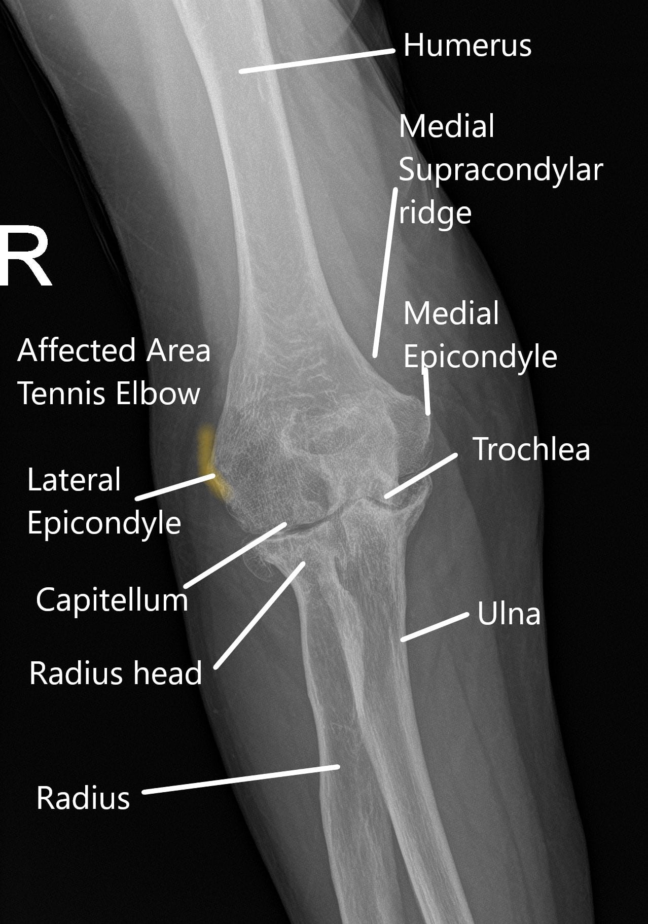
Tennis Elbow Joint Pain, Causes and Management Complete Orthopedics
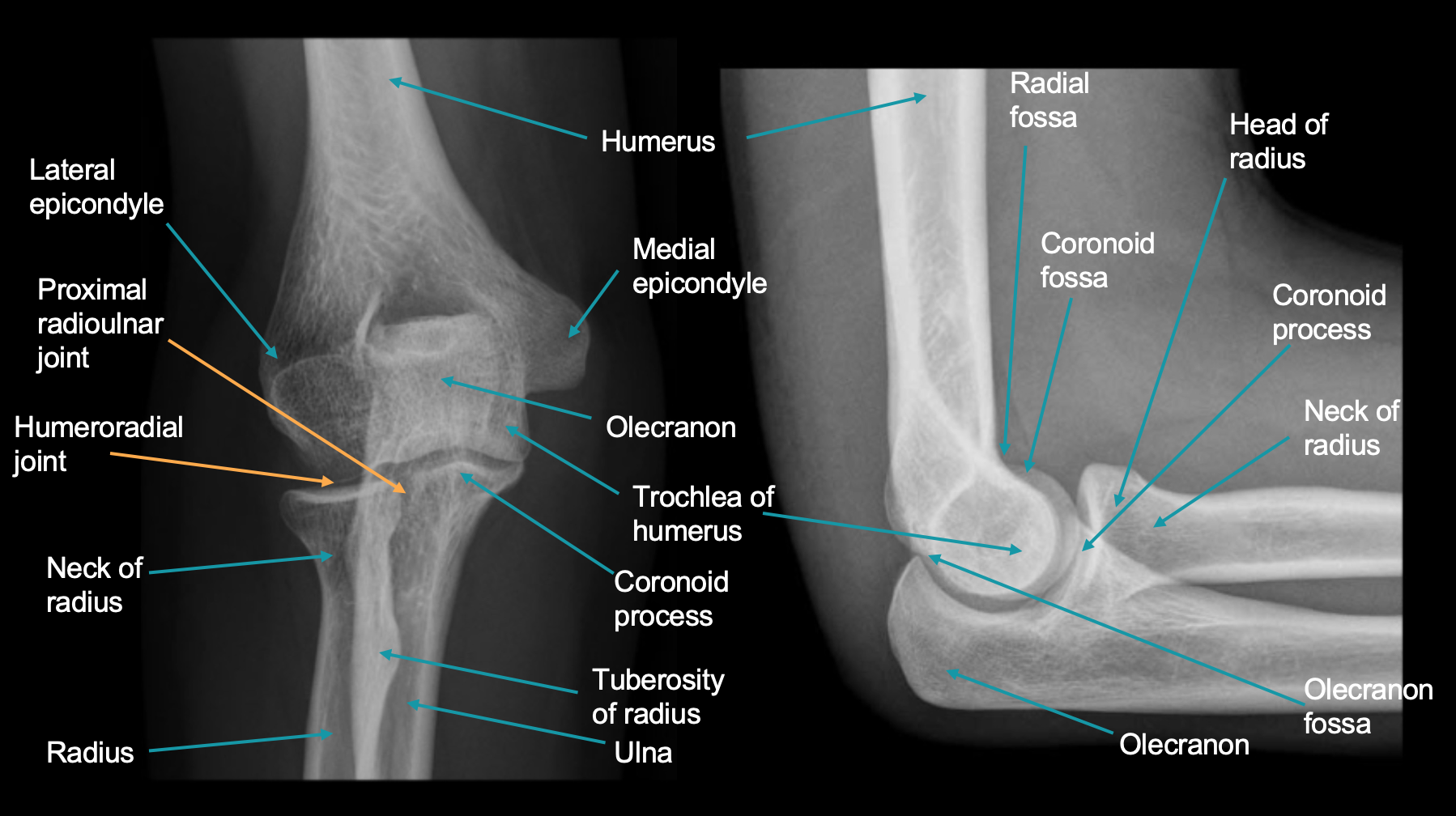
The radioanatomy of the elbow is studied via an AP X-Ray image and one in profile, showing the medial and lateral epicondyles, the olecranon, the head and neck of the radios, the radial and olecranon fossae, the humeral trochlea and the anatomical structures composing the humeroulnar joint, humeroradial joint and proximal radioulnar joint.

Typical pediatric elbow radiograph. The radiologic anatomy of a... Download Scientific Diagram
Medial Epicondyle Fracture: 5-10% of pediatric elbow fractures are medial epicondyle fractures . An important mechanism for developing this is elbow dislocation during which the medial epicondyle can be avulsed. During reduction, that small epicondyle can actually get stuck in the joint capsule and can easily be overlooked when reading an x-ray.

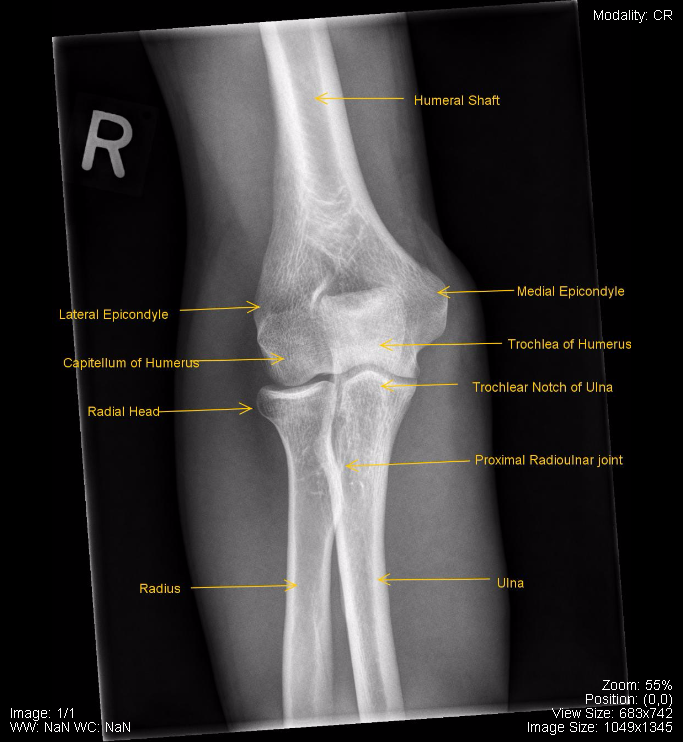
Elbow AP labelled
The sail sign On the lateral radiograph, inspect for the displacement of the anterior and posterior fat pads embedded in the two layers of the joint capsule. The anterior fat pad is normally seen as a faint line running with the distal humerus, whilst the posterior fat pad is not seen in normal radiographs.

Pediatric Elbow Anatomy in 2021 Elbow anatomy, Medical anatomy, Pediatrics
How to Read Emergency Images How to read an elbow x-ray Fractures lines can be difficult to visualize after acute elbow injury, particularly in children. Below are eight sequential steps to aid in the radiographic recognition of occult signs of injury.
