Scientists devised functioning kidney tissue

It's Okay To Be Smart Microscopic photography, Things under a
Contributions of electron microscopy to our knowledge of fine kidney structure have been reviewed and extended. Notable findings include the following: 1 . The glomerular capillary is extraordinarily specialized, presumably to facilitate diffusion. Its epithelial cells are highly branched, and have vast numbers of terminal processes that.

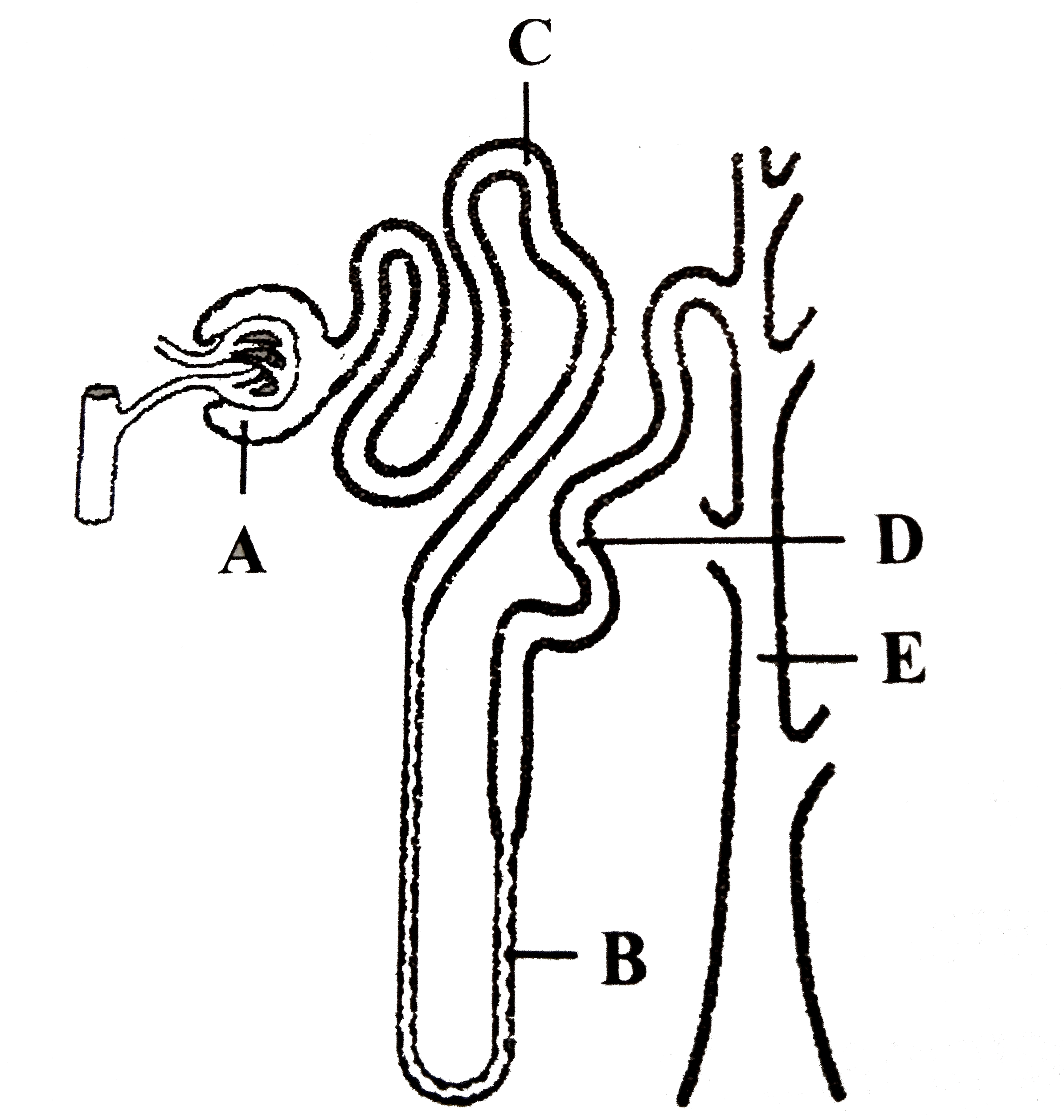
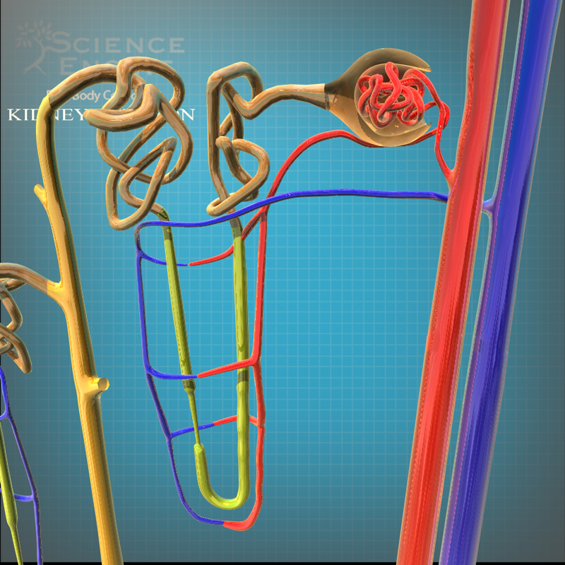
The nephron has five regions.
Understand the structure and function of the nephron and know the roles of the glomerulus, proximal tubule, loop of Henle, distal tubule, and collecting duct on urine formation and composition.. PAS stain, ×400) and wide foot process effacement (electron microscope, upper middle panel, ×2500). The second biopsy performed at age.

Nephron stock illustration. Illustration of anatomy, diuretics 47649327
To this purpose, we took advantage of scanning electron microscopy (SEM), an imaging approach rarely used for patients 15, which provides a global visualization of the actual three-dimensional.

Selfassembly of renal nephronlike tubules. (a) Fluorescence
75 of The Top 100 Retailers Can Be Found on eBay. Find Great Deals from the Top Retailers. Looking For Electron? We Have Almost Everything on eBay.

Electron microscope radioautographs of portions of the rat nephron
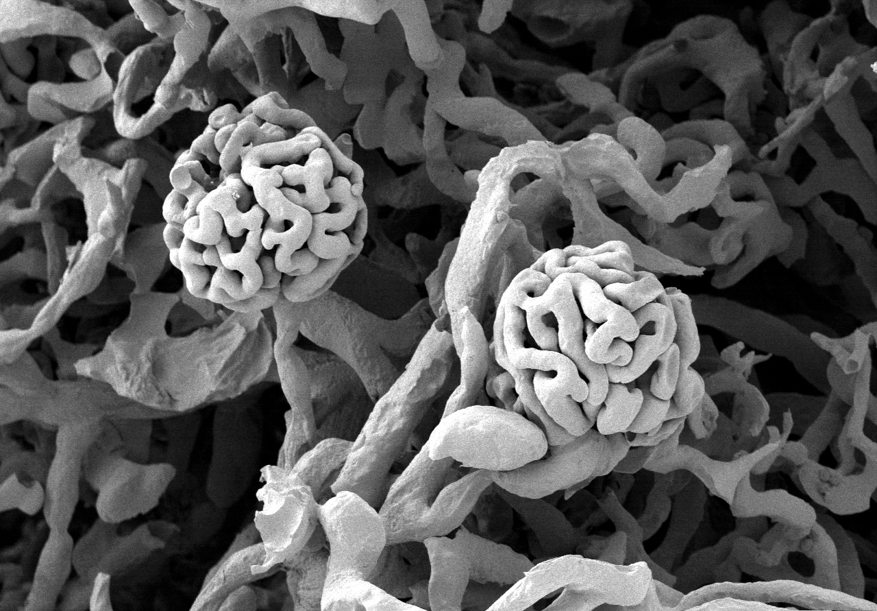
Scanning electron microscopy was used to study the ultrastructural morphology of the nephron. Material for observation was taken from rat kidneys which were fixed by vascular perfusion. Different techniques for splitting open the kidney, combined with stereoscopic viewing, provided many instructive views of nephron morphology.

nephron under microscope Diagram Quizlet
1/4. Synonyms: Cortex renalis. The kidneys are paired retroperitoneal organs of the urinary system. Their function is to filter blood and produce urine. Each kidney consists of a cortex, medulla and calyces. The nephron is the main functional unit of the kidney, in charge of removing metabolic waste and excess water from the blood.

The Urinary System Kidneys
Scanning electron microscopy revealed a number of new features including the complex organization and structure of kidney podocytes; the distribution of endothelial pores and the presence of endothelium microprojections and branching endothelial thickenings. Scanning electron microscopy was used to study the ultrastructural morphology of the nephron. Material for observation was taken from rat.
Structure Of Nephron Class 11 Jami of All Trades
The renal structures that conduct the essential work of the kidney cannot be seen by the naked eye. Only a light or electron microscope can reveal these structures. Even then, serial sections and computer reconstruction are necessary to give us a comprehensive view of the functional anatomy of the nephron and its associated blood vessels.

Nephrons in your kidneys Interior del cuerpo humano, Anatomia
The nephron is composed of the glomerulus, the juxtaglomerular complex, the proximal convoluted tubule, the loop of Henle and the distal convoluted tubule. This chapter deals with the light and electron microscopic structure of both the connective tissue capsule and the uriniferous tubule in the parenchyma of the mammalian kidney. Keywords
Scientists devised functioning kidney tissue
5 ELECTRON MICROSCOPY OF THE URINIFEROUS TUBULES* Jan L. E. Ericsson and Benjamin F. Trump I. II. III. INTRODUCTION FIXATION AND PREPARATION OF TISSUES FOR ELECTRON MICROSCOPY STRUCTURE OF THE MAMMALIAN TUBUI E 351 353 357 A. B. IV. Terminology Fine Structure Cyclostomes Teleosts Amphibia Reptiles Birds CORRELATION 357 358 416 STRUCTURE OF THE SUBMAMMALIAN VERTEBRATE TUBULE . .

Nephron illustration Medicine Notes, Medicine Studies, Renal Physiology
Panel c shows scanning electron microscopy-energy dispersive X-ray analyzer (SEM-EDX) mapping of the wafers. In the wafer shown (Na selective wafer), it is seen that the wafer before the run has a.
nephron anatomy 3d c4d
Microanatomy of the Nephron Renal Corpuscle. As discussed earlier, the renal corpuscle consists the glomerulus and the glomerular capsule. The glomerulus is a high pressured, fenestrated capillary with large holes (fenestrations) between the endothelial cells.The glomerular capsule captures the filtrate created by the glomerulus and directs this filtrate to the PCT.
Transmission electron microscopy showing the presence of specific
Download the Temu App and start saving more today! Unleash incredible deals and coupons. Ready to shop and save? Explore amazing deals on the Temu App. Free shipping & return.
Scanning Electron Microscopy of Corrosion Casts
The renal structures that conduct the essential work of the kidney cannot be seen by the naked eye. Only a light or electron microscope can reveal these structures. Even then, serial sections and computer reconstruction are necessary to give us a comprehensive view of the functional anatomy of the nephron and its associated blood vessels.
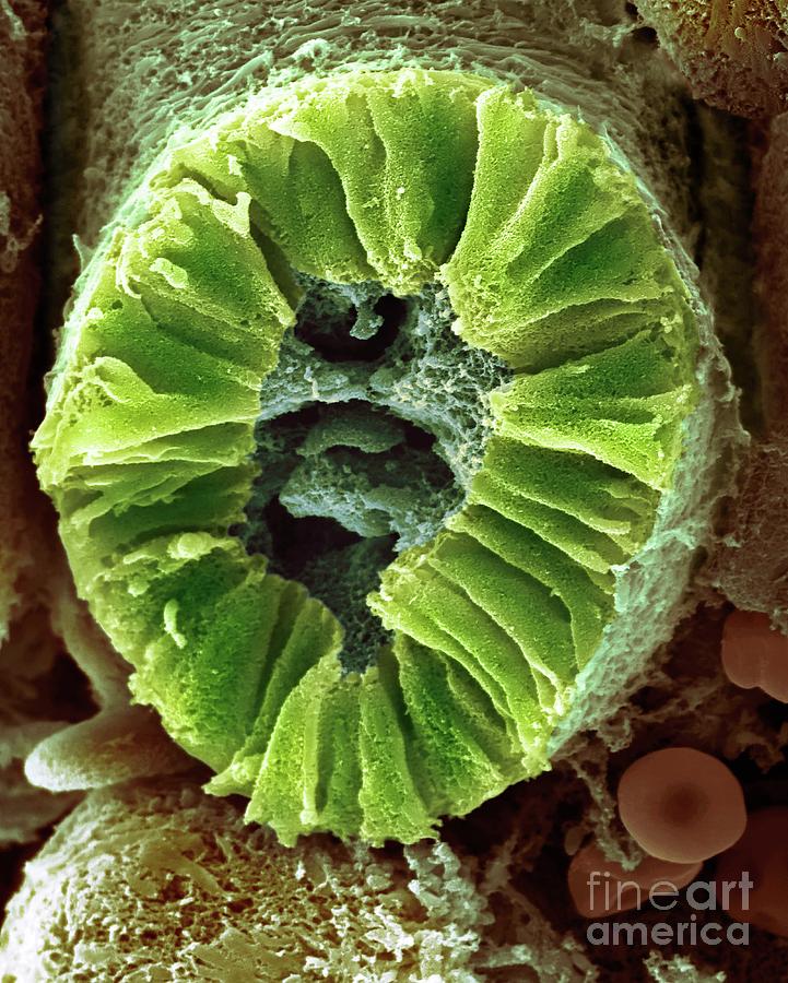
Human Kidney Nephron Photograph by Dennis Kunkel Microscopy/science
Symposium on Renal Physiology Electron Microscopy of the Kidney' JOHANNES RHODIN, M.D. New York, New York THE structure of the nephron includes a great variety of cell types, from the complicated composition of the filtering glomerular capillary membrane to the relatively simple and pale cells of the collecting ducts.
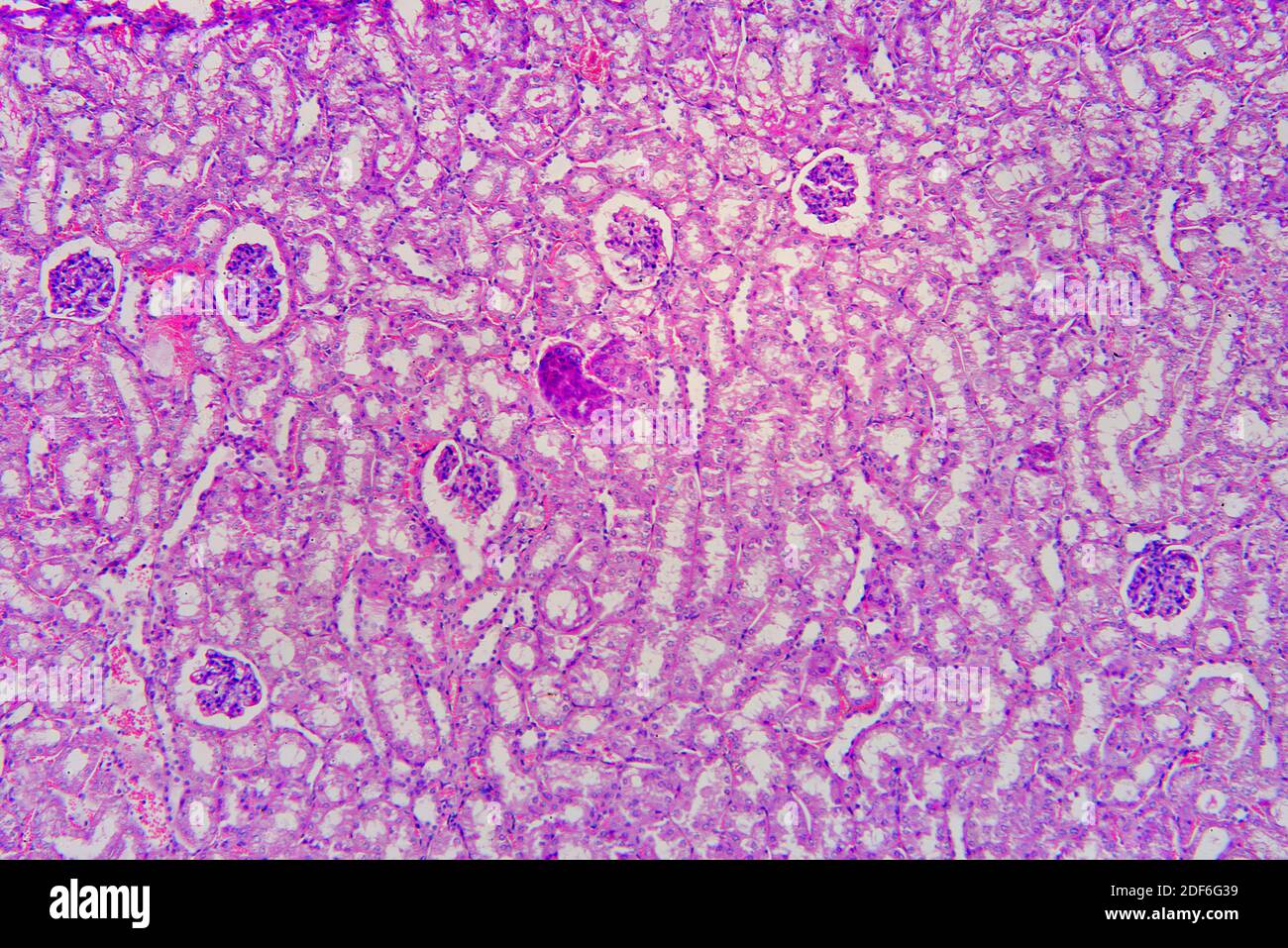
Kidney section showing nephrons, Bowman capsules, glomerulus and distal
Scanning electron microscopy was used to study the ultrastructural morphology of the nephron. Material for observation was taken from rat kidneys which were fixed by vascular perfusion. Different techniques for splitting open the kidney, combined with stereoscopic viewing, provided many instructive views of nephron morphology. In addition.
