Olympus BX51 Phase Contrast Microscope Darkfield & Phase Microscope Central

Olympus BX51 Phase Contrast Microscope Darkfield & Phase Microscope Central
Darkfield microscopy is a simple and popular method for rendering unstained and transparent specimens clearly visible. Good candidates for darkfield observation often have refractive indices very close in value to that of their surroundings and are difficult to image with conventional brightfield techniques.

40X2000X Trinocular Compound Darkfield Microscope with Oil Condenser Microscope Central
Dark-field microscopy is a technique that can be used for the observation of living, unstained cells and microorganisms. In this microscopy, the specimen is brightly illuminated while the background is dark. It is one type of light microscope, others being bright-field, phase-contrast, differential interface contrast, and fluorescence.
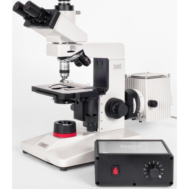
Hund H 600 LL 100 HP dark field microscope
The surface beneath the opening should be a flat black. Turn off any built-in illuminator. Aim a high-intensity light source toward the specimen at an angle, from the top or side through a glass dish or jar. With a compound microscope, dark field is obtained by placing an occulting disk in the light path between source and condenser.

What is a Dark Field Microscope? (with pictures)
Dark field Microscopy, also known as dark field illumination, is a method that uses microscopes to make a specimen appear bright white. This effect is achieved by removing dispersed light so that the specimen is appearing through scattered light only. Doing this produces a bright white image on a dark background.
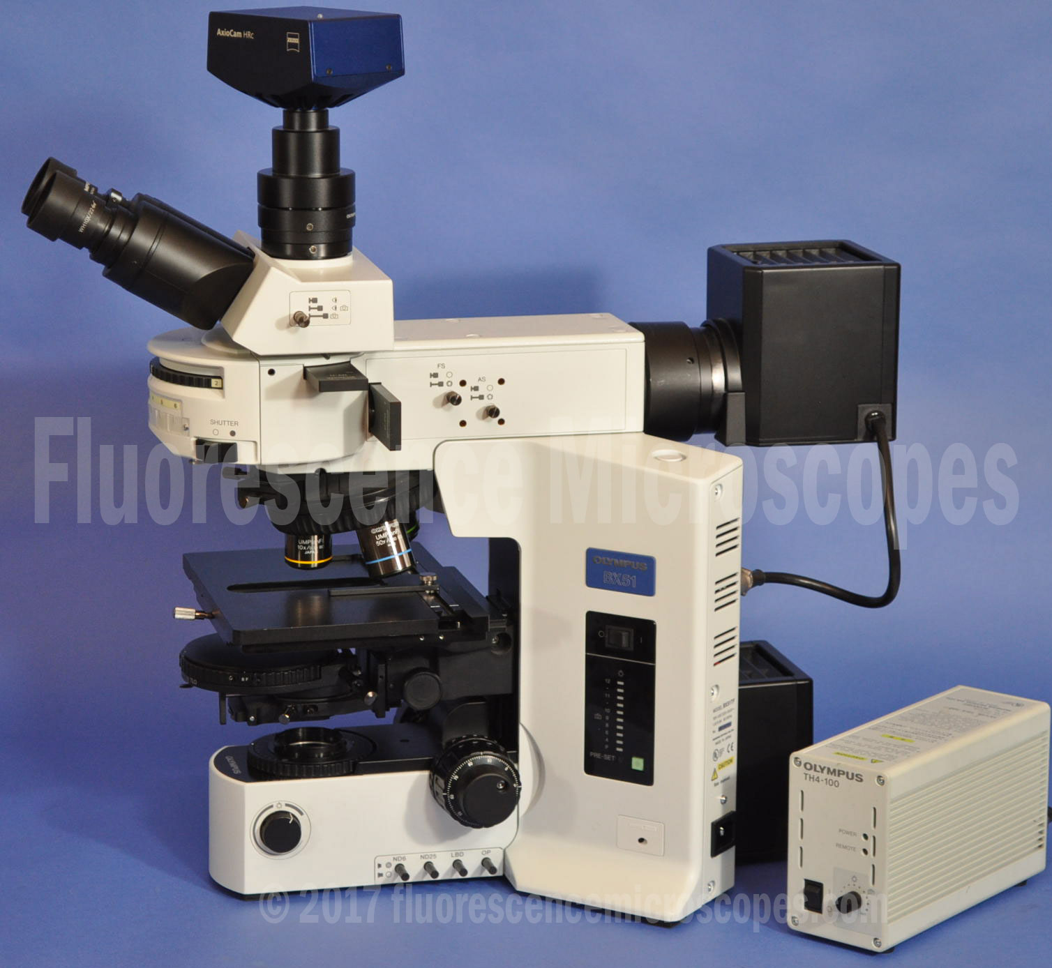
Olympus BX51 Upright DIC Darkfield Metallurgical Microscope
Dark-field X-ray microscopy ( DFXM [1] or DFXRM [2]) is an imaging technique used for multiscale structural characterisation. It is capable of mapping deeply embedded structural elements with nm-resolution using synchrotron X-ray diffraction -based imaging.
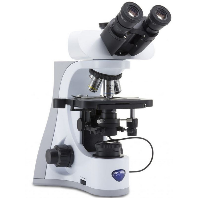
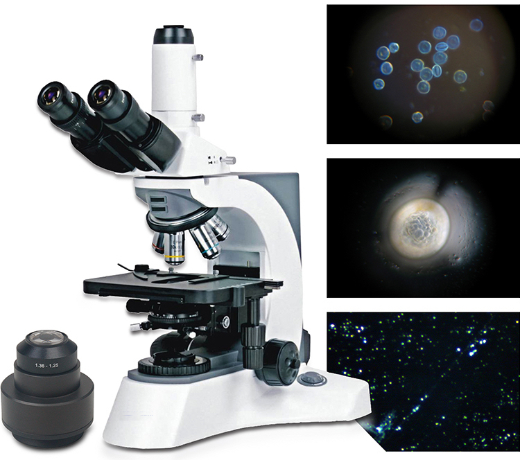
Optika B510DK, darkfield, trinocular microscope, 1000x, IOS, for live blood analysis
What is darkfield microscopy? What are its key advantages? Learn everything you need to know about imaging with darkfield in this blog post.

Nikon 50i Darkfield Microscope Microscope Central
What is Darkfield Microscopy? How does Darkfield Microscopy work? What types of specimens are suitable for Darkfield Microscopy? What are the advantages of Darkfield Microscopy?

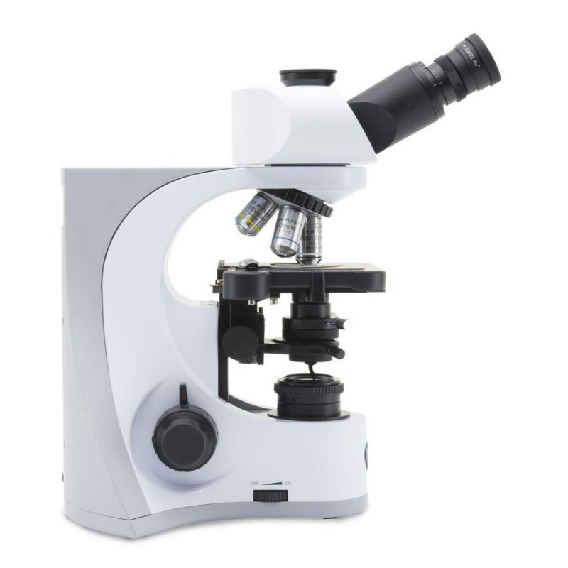
Olympus BX40 Phase Contrast & Darkfield Microscope Microscope Central
Dark-field microscopy (also called dark-ground microscopy) describes microscopy methods, in both light and electron microscopy, which exclude the unscattered beam from the image. Consequently, the field around the specimen (i.e., where there is no specimen to scatter the beam) is generally dark.
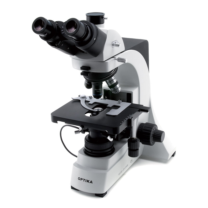
Optika B 500TDK trinocular darkfield microscope
Dark-field microscopy gains popularity as a background-free detection tool, where small, bright features appear on a dark background.

Nikon 50i Darkfield Microscope Microscope Central
A darkfield microscope is a compound microscope which uses an aperture in the shape of an annulus that is placed between the light source and condenser lens. Ring-shaped light that passes through the aperture is focused by the condenser onto the biological specimen or material sample to be observed.

BDSW3001 dark field trinocular Biological MicroscopeShenzhen Boshida Optical Itrument Co., Ltd.
Dark-field microscopy is slightly more complicated in stereomicroscopy. A specialized stand that has a reflection mirror and a plate to shield light is required. This directs an inverted hollow.
Optika B510DK, darkfield, trinocular microscope, 1000x, IOS, for live blood analysis
Dark-field microscopy is ideally used to illuminate unstained samples causing them to appear brightly lit against a dark background. This type of microscope contains a special condenser that scatters light and causes it to reflect off the specimen at an angle. Rather than illuminating the sample with a filled cone of light, the condenser is.
BUM500DFL Digital Dark field Laboratory Microscope
This principle is applied in darkfield (also called ) microscopy, a simple and popular method for making transparent specimens clearly visible. Such objects often have refractive indices very close in value to that of their surroundings and are difficult to image in conventional brightfield microscopy.
Dark Field Microscope HDSet SARRAS e.U.
Chapter 9 Dark Field Microscopy. C. Robert Bagnell, Jr., Ph.D., 2012. In the 1840's, the ultimate test objects for light microscopes were diatoms, in particular the species then known as Navicula Spencerii. This diatom was named after Charles Spencer, the New York lens maker whose lenses could resolve this diatom's striae.
Darkfield Microscope AmScope Supplies Trinocular Darkfield Compound Biological Microscope with
What is dark field microscopy? Dark field microscopy is a type of microscopy technique that is used in both light and electron microscopy, where only the specimen is lit by a light or electron beam, and the rest of the specimen field is dark.

Motic Panthera TEC Metallurgical Brightfield / Darkfield Microscope Microscope Central
A dark field microscope operates on the opposite principle of a brightfield microscope, and the entire field of view appears dark when there is no specimen and once a specimen is placed, it appears bright over this dark background. Hence the name dark field microscope. Table of Contents What is Dark field microscope Working Principle and Diagram
