Diagrams of Microscope 101 Diagrams
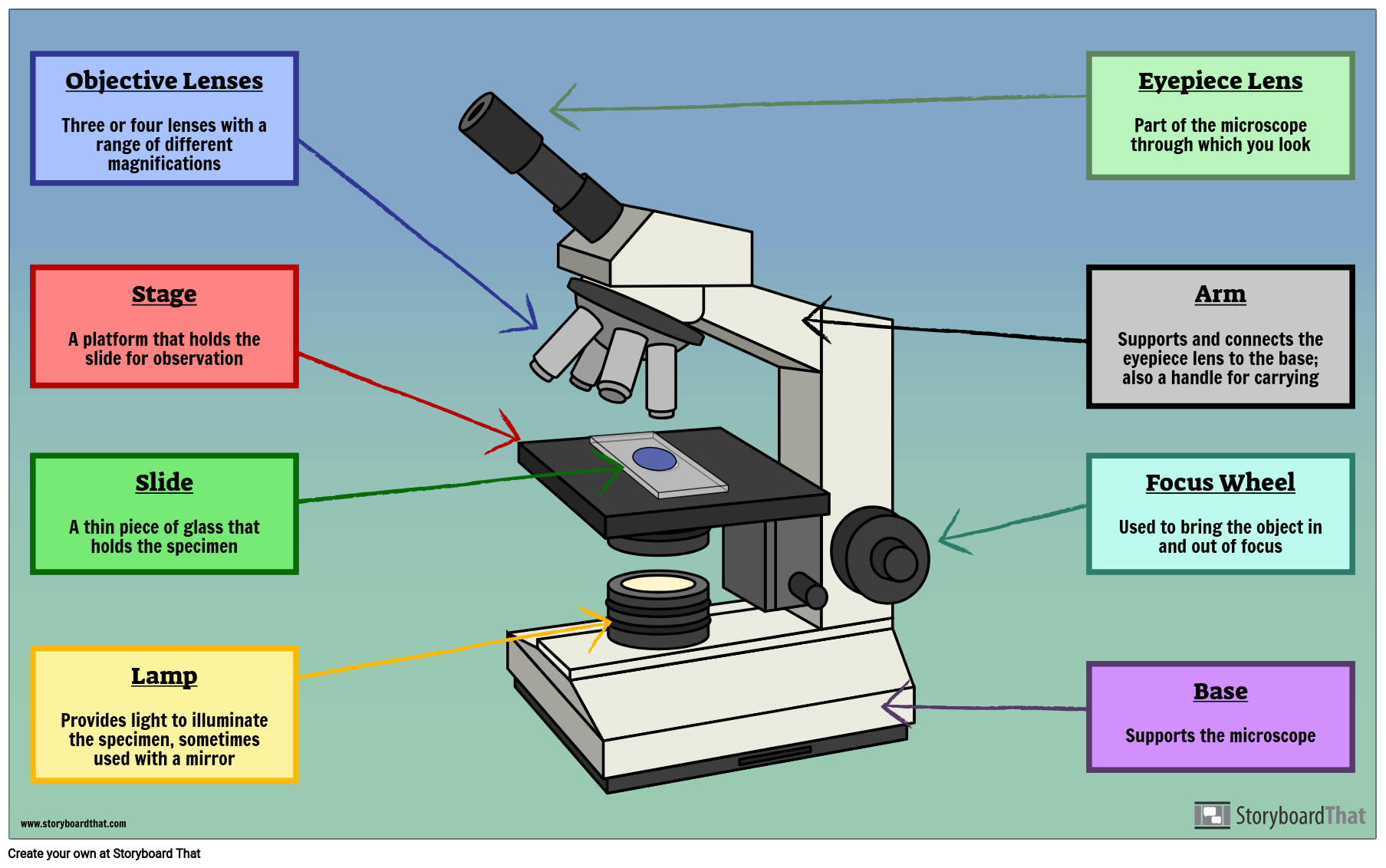
Labelled Microscope with Functions Storyboard by oliversmith
There are two major types of electron microscopy. In scanning electron microscopy ( SEM ), a beam of electrons moves back and forth across the surface of a cell or tissue, creating a detailed image of the 3D surface. This type of microscopy was used to take the image of the Salmonella bacteria shown at right, above.
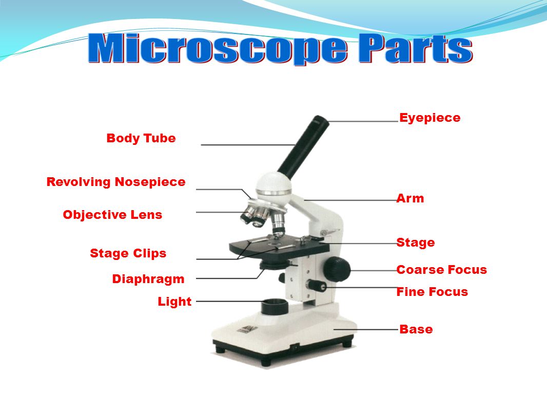
Monday September 25 Parts of a Compound Light Microscope
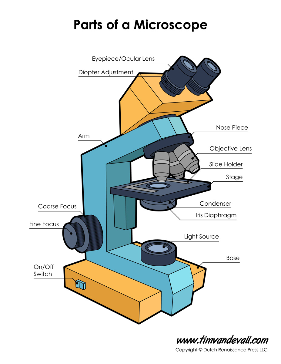
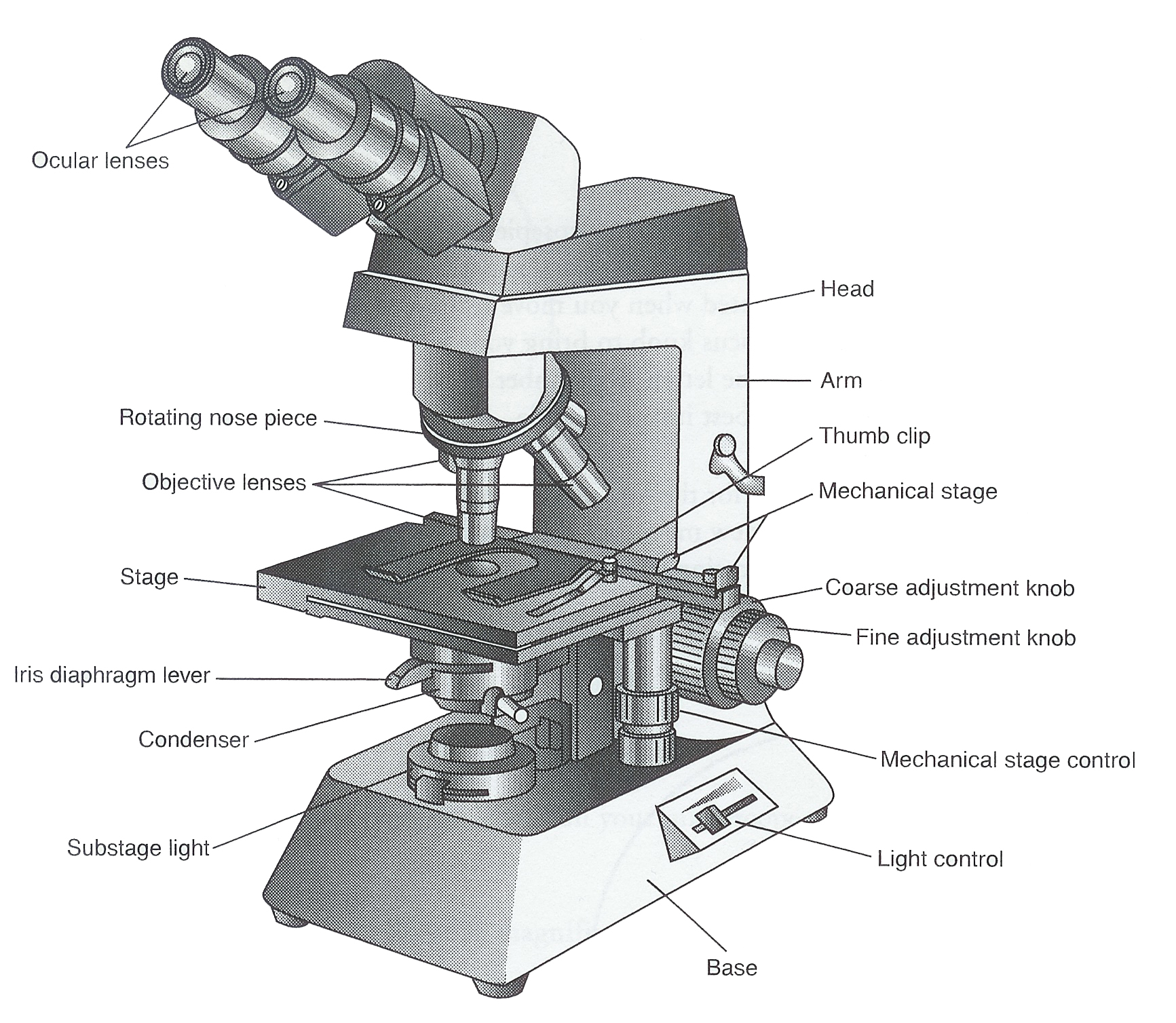
Tube: Connects the eyepiece to the objective lenses. Arm: Supports the tube and connects it to the base. Base: The bottom of the microscope, used for support. Illuminator: A steady light source (110 volts) used in place of a mirror. If your microscope has a mirror, it is used to reflect light from an external light source up through the bottom.

Microscope Diagram to Print 101 Diagrams
Simple microscope is a magnification apparatus that uses a combination of double convex lens to form an enlarged, erect image of a specimen. The working principle of a simple microscope is that when a lens is held close to the eye, a virtual, magnified and erect image of a specimen is formed at the least possible distance from which a human eye.

Microscope Diagram Labeled, Unlabeled and Blank Parts of a Microscope
So, a compound microscope with a 10x eyepiece magnification looking through the 40x objective lens has a total magnification of 400x (10 x 40). Specimen or slide: The object used to hold the specimen in place along with slide covers for viewing.. Compound Microscope Parts, Functions, and Labeled Diagram. Parts of a Compound Microscope.
Compound Microscope Parts Labeled Diagram and their Functions Rs
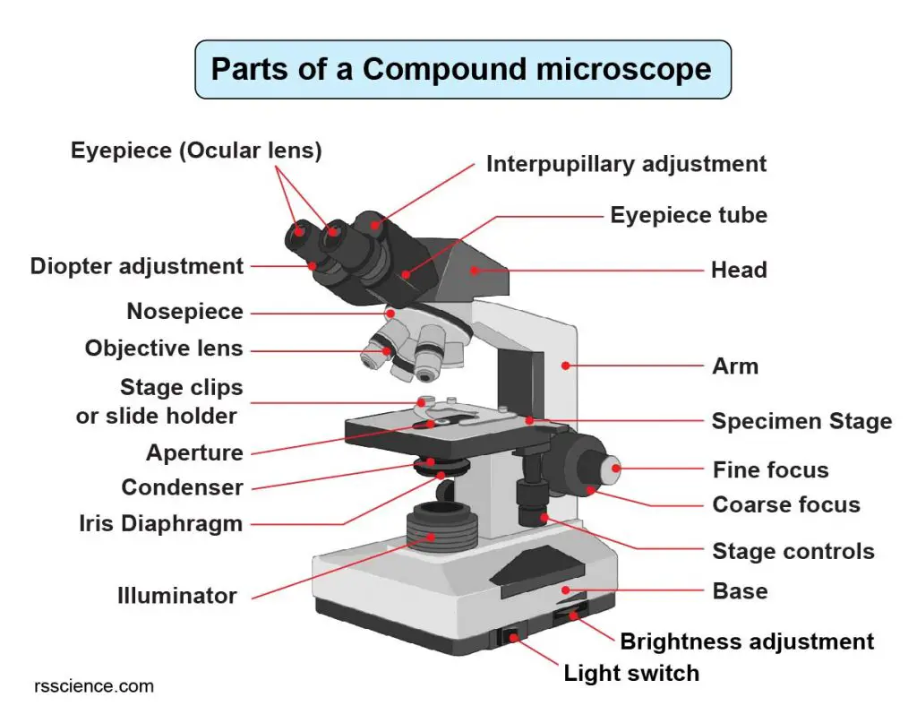
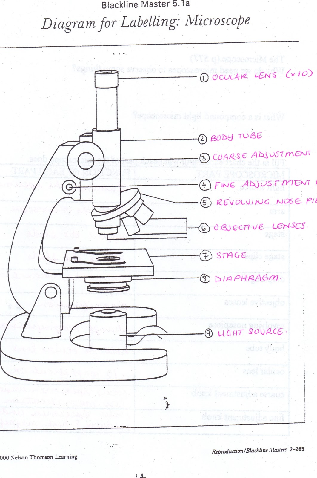
Figure: Diagram of parts of a microscope. There are three structural parts of the microscope i.e. head, arm, and base. Head - The head is a cylindrical metallic tube that holds the eyepiece lens at one end and connects to the nose piece at other end. It is also called a body tube or eyepiece tube.
Ag Biology Unit 2
Microscope Parts and Functions With Labeled Diagram and Functions How does a Compound Microscope Work?. Before exploring microscope parts and functions, you should probably understand that the compound light microscope is more complicated than just a microscope with more than one lens.. First, the purpose of a microscope is to magnify a small object or to magnify the fine details of a larger.

Parts of a Compound Microscope Labeled (with diagrams) Medical
Labeling the Parts of the Microscope This activity has been designed for use in homes and schools. Each microscope layout (both blank and the version with answers) are available as PDF downloads. You can view a more in-depth review of each part of the microscope here. Download the Label the Parts of the Microscope PDF printable version here.

How to Use a Microscope (Properly) Step by Step New York Microscope
The web page titled "Parts of a Microscope with Labeled Diagram and Functions" has the following key takeaways: 🔍 The microscope is an essential tool for scientists, researchers, and medical professionals. 🧬 The main function of a microscope is to provide a magnified view of small objects or organisms, such as bacteria, cells, or tissues.
Microscopes 7th Grade Science
Eyepiece lens magnifies the image of the specimen. This part is also known as ocular. Most school microscopes have an eyepiece with 10X magnification. 2. Eyepiece Tube or Body Tube. The tube hold the eyepiece. 3. Nosepiece. Nosepiece holds the objective lenses and is sometimes called a revolving turret.

Light Microscope Definition, Principle, Types, Parts, Labeled Diagram
Phase-contrast microscope labeled diagram. Phase-contrast microscope functions: Its applications areas include. In cases where the specimen is colorless and is very tiny; In biology to conduct cellular level examination of microorganisms that can't be visualized using the bright field microscopy; Interference Microscope
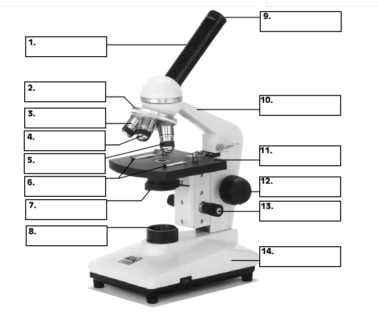
All Saints Online Diagram for Labelling Microscope
In this interactive, you can label the different parts of a microscope. Use this with the Microscope parts activity to help students identify and label the main parts of a microscope and then describe their functions. Drag and drop the text labels onto the microscope diagram. If you want to redo an answer, click on the box and the answer will.

Parts of a microscope with functions and labeled diagram
A Study of the Microscope and its Functions With a Labeled Diagram To better understand the structure and function of a microscope, we need to take a look at the labeled microscope diagrams of the compound and electron microscope. These diagrams clearly explain the functioning of the microscopes along with their respective parts.

Microscope Diagram to Print 101 Diagrams
Fluorescence microscopes: These use fluorescent dyes to highlight specific structures or molecules in a sample and are commonly used in biological research. X-ray microscopes: These use X-rays to produce images of the internal structure of samples and are often used to study materials and biological specimens.

Microscope labeled diagram
A labeled diagram of microscope parts furnishes comprehensive information regarding their composition and spatial arrangement within the microscope, enabling researchers to comprehend their function effectively. In this comprehensive article, we will delve into the intricate parts of the microscope, exploring their functions in detail..
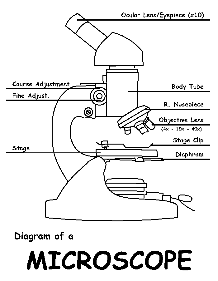
Diagram of a Microscope by ScienceDoodles on DeviantArt
A light microscope is a biology laboratory instrument or tool, that uses visible light to detect and magnify very small objects and enlarge them. They use lenses to focus light on the specimen, magnifying it thus producing an image. The specimen is normally placed close to the microscopic lens.
Microscope Parts Drawing at GetDrawings Free download
The description given below summarize the brief description of microscope parts used to visualize the microscopic specimens such as animal cells, plant cells, microbes, bacteria, viruses, microorganisms etc. The Microscopes parts divided into three different structural parts Head, Base, and Arms. Head/Body: It contain the optical parts in the.
